Lymphogranuloma Venereum
26
AT-A-GLANCE
■ Lymphogranuloma venereum is a sexually transmitted infection caused by L serovars (serologic variants) of Chlamydia trachomatis.
■ Endemic in Africa, Southeast Asia, and South and Central America, and rare in developed countries.
■ Outbreaks have occurred among men who have sex with men in Europe, Australia, and North America.
■ Clinically manifests as inguinal and anorectal syndromes, in 3 stages.
■ Hematogenous spread with manifestations of systemic infection.
■ Diagnosis is by identification of organism and by serology or genotyping.
■ Doxycycline or azithromycin (second-line) treatment is curative if given early in the infection course.
EPIDEMIOLOGY
Lymphogranuloma venereum (LGV) is a sexually transmitted infection caused by specific Chlamydia variants that is rare in developed countries. It is endemic in East and West Africa, India, Southeast Asia, South and Central America, and some Caribbean Islands; LGV accounts for 7% to 19% of genital ulcer diseases in areas of Africa and India.1,2 The peak incidence occurs in sexually active persons 15 to 40 years of age, in urban areas, and in individuals of lower socioeconomic status. Men are 6 times more likely than women to manifest clinical infection.3 The incidence of LGV is low in the developed world where cases are usually limited to travelers or military personnel returning from endemic areas. Since 2003, however, outbreaks of LGV have appeared in Europe, Australia, and North America, particularly in the form of proctitis, among HIV-positive men who have sex with men (MSM).4-8 In 2013, more than 1000 cases were reported to the European Centre for Disease Prevention and Control, approximately 50% of them from the United Kingdom9; however, the real number is likely to be higher, as LGV is not routinely recorded in many European countries. In addition, a cluster of 38 LGV cases among MSM was reported in the United States in Michigan for the period August 2015 thru April 201610; the true incidence of LGV in the United States, however, is unknown because national reporting of LGV ended in 1995.
LGV is contracted by direct contact with infectious secretions, usually through any type of unprotected intercourse, whether oral, vaginal, or anal. Transmission efficiency is unknown.11 Asymptomatic rectal infection and/or penile and oral infection is the likely source of onward transmission.12 Sexual practices, such as fisting and sex-toy sharing, may be other routes of transmission. In a study that compared sexual behaviors in men with LGV and men with non-LGV chlamydial proctitis, fisting was a major predisposing factor.13 An epidemic of LGV has been reported among “crack” cocaine users in the Bahamas.14
As a consequence of underdiagnosis and underreporting, the epidemiology of LGV remains poorly understood. Common diagnostic laboratory methods are nonspecific and not readily available in endemic areas. Even in industrialized countries, only a few laboratories offer specific assays to LGV serovars (serologic variants).15 Without such assays, many LGV cases are misdiagnosed as common chlamydial urogenital infection. Underdiagnosis of LGV is also largely a result of the presence of an asymptomatic carrier state. Women, in particular, may harbor asymptomatic persistent infection in the cervical epithelium, thus serving as reservoirs of the infection as they do for other urogenital chlamydial infections and gonorrhea. A large study conducted in the United Kingdom found that only 6% of MSM were asymptomatic carriers of LGV Chlamydia serovars; the majority of cases of LGV in the rectum and urethra were symptomatic,16 whereas a recent study reported a prevalence of asymptomatic rectal infection of 19%.17 Infectivity in men usually ceases after healing of the primary mucosal lesion. Interestingly, most of the detected cases in the recent outbreaks are in men who practice receptive anal sex, suggesting that a high proportion of men who practice insertive anal sex are misdiagnosed or undiagnosed. The reasons behind this are unclear but may be organismrelated, host-related (eg, sexual practices such as fisting and the use of sex toys, IV drug use, HIV status) or physician-related (failure to diagnose genital LGV). The resurgence of LGV as a health problem may simply reflect increased awareness rather than increased incidence.
ETIOLOGY AND PATHOGENESIS
LGV (tropical or climatic bubo, lymphopathia venerea, Nicolas-Favre disease) is caused by Chlamydia trachomatis serovars L1, L2, and L3.18 Most of the recent outbreaks are caused by a number of strains of
26
C. trachomatis L2 serovar (L2b), suggesting that these outbreaks most likely represent increased awareness of a slowly evolving endemic.5,6,16,19. There are new cases in Spain, Finland, Czech Republic, and The Netherlands (where the first case of a female patient with bubonic LGV caused by a serovar L2b was reported).20-23 In France, a change in epidemiology since 2013 has been reported with declining infections of the L2b type and increasing numbers of the L2 type.24 Case reports with infection of the L2c variant suggest a more aggressive clinical course, causing severe proctitis.25
Chlamydiae are obligate intracellular bacteria characterized by 2 distinct morphologic forms: (a) the small metabolically inactive and infectious elementary body, and (b) the larger metabolically active and noninfectious reticulate body (Fig. 173-1). C. trachomatis has been subdivided into several serovars that differ in their major outer membrane proteins, and which are associated with several diseases. Serovars A, B, and C are the causes of trachoma ocular infections. These infections are rarely seen in North America and Europe, but are quite frequently seen in Africa and Asia, where they represent an important cause of blindness.26 Serovars D to K are responsible for ocular, genital, and respiratory tract infections.15,27,28 In contrast to the A-to-K serovars that remain confined to the mucosa, LGV serovars are more invasive and have a high affinity for macrophages.18 After being inoculated onto the mucosal surface, the organisms replicate within macrophages, and find their way to the draining lymph nodes, and cause lymphadenitis.
CLINICAL FINDINGS
CUTANEOUS LESIONS AND RELATED PHYSICAL FINDINGS
CUTANEOUS LESIONS
AND RELATED PHYSICAL
FINDINGS
Clinical manifestations are protean, depending on the sex of the patient, acquisition mode, and the infection stage. Three clinical stages characterize LGV. Nonspecific cutaneous lesions such as erythema nodosum,
3194
erythema multiforme, urticaria, and scarlatiniform exanthema may occur with any of these stages.29
PRIMARY STAGE
Three to 30 days after infection, 5- to 8-mm painless erythematous papule(s) or small herpetiform ulcers appear at the site of inoculation (Fig. 173-2). Painful ulcerations30 and nonspecific urethritis are less common. Thus, the initial lesions may be differentiated from the more common herpetic lesions by the lack of pain associated with the lesions. In males, the lesion is usually found on the coronal sulcus, prepuce, or glans penis, and in females, on the posterior wall of the vagina, vulva, or, occasionally, the cervix. Inoculation also may be rectal, at the lip, or pharyngeal.15,31 The primary lesion is transient, often heals within a few days, and may go unnoticed.25
SECONDARY STAGE
A few weeks after the primary lesion appears, marked lymph node involvement and hematogenous dissemination occur, manifested by variable signs and symptoms, including fever, myalgia, decreased appetite, and vomiting. Photosensitivity may develop in up to 35% of the patients, often 1 to 2 months after bubo formation (painful inflammation of lymph node, characterized by a unilateral enlargement, suppuration, and abscesses, also firm and tender immovable mass).32 Less commonly, patients may develop meningoencephalitis, hepatosplenomegaly, arthralgia, and iritis.33,34 The lymphadenitis episodes often resolve spontaneously in 8 to 12 weeks. Depending on the mode of transmission, 2 major syndromes are seen.
The acute genital syndrome or inguinal syndrome is characterized by inguinal and/or femoral lymph node involvement and is the major presentation in men in developing countries. Initially, the skin overlying the affected lymph node is erythematous and indurated (Fig. 173-3). Over the subsequent 1 to 2 weeks, the lymph node enlarges and coalesces to form a firm and tender immovable mass (bubo; Fig. 173-4), which may rupture and drain through the skin, forming sinus tracts. Bilateral involvement occurs in onethird of the cases (Fig. 173-5). Nodal enlargement on either side of the inguinal ligament, the “groove sign,” is pathognomonic of LGV, but only presents in 10% to 20% of cases35 and is rarely bilateral.15 In women, inguinal lymphadenitis is unusual because the lymphatic
26
drainage of the vagina and cervix is to the deep pelvic/ retroperitoneal lymph nodes. When these nodes are involved, low abdominal/back pain that exacerbates upon lying supine and pelvic adhesions may ensue. The acute anorectal syndrome is characterized by perirectal nodal involvement, acute hemorrhagic proctitis, and pronounced systemic symptoms. It is the most common presentation in women and in homosexual men who practice anal sex. The major source of rectal spread in women is the internal lymphatic drainage of the lower two-thirds of the vagina. Patients may complain of anal pruritus, bloody and/or purulent rectal discharge, tenesmus, diarrhea, constipation, and lower abdominal pain.36 In a recent outbreak of LGV in Western Europe, 96% of MSM patients presented with signs and symptoms of proctitis.37 LGV proctitis mimics chronic inflammatory bowel disease, both clinically and in pathologic substrate. These cases may present with an incomplete or undisclosed history of proctosigmoiditis, without the characteristic adenopathy syndrome.38 Reactive arthritis in MSM following LGV proctitis has been reported in several cases in recent years.34 Despite affecting mostly HIV-positive MSM, LGV has not behaved as an opportunistic infection in the recent outbreak, and clinical features have not differed between HIV-positive and HIV-negative cases.39
TERTIARY STAGE
This stage is seen more often in women with untreated anorectal syndrome than in men, although it is also seen in homosexual men, because of the location of the involved lymphatics. It includes rectal strictures (most common) and abscesses, perineal sinuses, rectovaginal fistulae (leading to “watering can perineum”), and “lymphorrhoids” (perianal outgrowths of lymphatic tissue). Esthiomene (Greek for “eating away”) is a rare primary infection of the external genitalia (mostly in women), leading to progressive lymphangitis and genital destruction. Infertility and “frozen pelvis” are potential sequelae of ruptured deep pelvic nodes in women. Late sequelae of the genital syndrome are less common and include urethral strictures and
3195
26
genital elephantiasis with ulcers and fistulas (in 4% of patients).40 Penile deformities, such as the saxophone penis, may also occur.41
OTHER UNUSUAL MANIFESTATIONS
OTHER UNUSUAL
MANIFESTATIONS
Extragenital-anal inoculation of LGV is rare. Oropharyngeal infection may manifest initially as pinheadsized vesicles on the lip, and later on as cervical lymphadenopathy with constitutional symptoms, closely mimicking lymphoma. Tonsillitis, supraclavicular and mediastinal lymphadenopathy, and pericarditis rarely occur.42-44 Ocular autoinoculation of infected discharges may lead to conjunctivitis with marginal corneal perforation, often with preauricular lymphadenopathy.45 Although most rectal LGV among MSM appears to be symptomatic, some investigators have noted that asymptomatic infections can occur.46
LABORATORY TESTS
Diagnosis of LGV may be difficult, but LGV should be suspected in any patient with infected sexual contacts, genital ulcer, perianal fistula, or bubo. The accuracy of clinical diagnosis has been reported to be as low as 20%.47 Consequently, laboratory tests are important to establish the diagnosis and are usually divided into 2 broad categories: (a) nonspecific tests that do not distinguish between LGV and non-LGV serovars, and (b) specific LGV tests. In practice, a positive test on lymph node aspirate is considered diagnostic of LGV, in contrast to a positive test on a primary genital or anorectal lesion where further specific testing is required to rule out common chlamydial urogenital infections.
SPECIFIC TESTS FOR LYMPHOGRANULOMA VENEREUM
SPECIFIC TESTS FOR
LYMPHOGRANULOMA
VENEREUM
Confirmation of LGV requires identification of the genotype L1, L2 or L3. Typing is important because the recommended antibiotic treatment for LGV is longer than for non-LGV cases.25,48 Nucleic acid amplification tests (NAAT), like polymerase chain reaction (PCR), may be performed on all specimens, and has been the diagnostic method of choice in recent outbreaks. Several currently available commercial NAATs allow sensitive and specific detection of C. trachomatis, but do not provide any information about the underlying genotype(s).49,50 Using these tests identification of LGV in positive specimens would require a separate analysis. Methods for typing include genotype-specific PCRs, multiplex-nested PCR and RFLP- or sequence
3196
analysis of appropriate omp1 gene regions.51-54 LGV- and non LGV strains can also be differentiated by real time PCR tests based on pmpH or omp1 gene regions that contain specific deletions or single nucleotide polymorphisms.53,55,56
A real-time quadriplex PCR assay has been developed that is capable of detecting LGV, non-LGV, or mixed infections simultaneously in rectal specimens. The assay also contains a supplemental amplification target for the confirmation of C. trachomatis infection as well as a human DNA control for monitoring sample adequacy and PCR inhibition.55
Another assay described by Twin et al.56 targets a 61-bp PCR fragment of the omp1 gene, where LGV and non LGV serovars deviate from each other by one nucleotide that allows differentiation by high resolution melting analysis (HRMA). The assay correctly identifies LGV and non LGV types in 44/47 (93,6%) specimens previously analyzed by a multiplex real time PCR assay published from the same group.54
A dual target PCR assay based on the cryptic plasmid for highly sensitive detection of C. trachomatis and pmpH to differentiate between LGV and non LGV strains in a single reaction has been shown to distinguish LGV and non LGV infection in 95% of 156 analyzed C. trachomatis positive samples, including 65/67 (97%) anorectal smears.57 The advantage of this test is to use a second target recommended to prevent missed infections due to target sequence variation49 and to differentiate LGV and non-LGV genotypes simultaneously in a single reaction. All these assays are commercially unavailable inhouse developments that are not FDA approved. Nevertheless, these tests may be used for laboratory testing after evaluation of test performance characteristics according to quality assurance of microbiological diagnostics, such as CLIA (Clinical Laboratory Improvement Amendments).15
NONSPECIFIC CHLAMYDIAL TESTS
NONSPECIFIC
CHLAMYDIAL TESTS
The complement fixation test is the most commonly used test. Titers greater than 1:64 are considered diagnostic, titers greater than 1:256 are highly suggestive of LGV, and titers below 1:32 exclude the diagnosis unless the infection is in its early stages.58 The microimmunofluorescence test for the L-type serovar is more sensitive and specific, but less readily available.59 The use of species-specific proteins and peptides in immunoblots or line assays further improves the specificity of serologic testing,60 but as of this writing no LGV-specific serologic marker has been identified. In addition, direct fluorescence microscopy using conjugated monoclonal antibody against C. trachomatis on smears from bubo material or genital swab can be done.61 Serology assays are sensitive but nonspecific because of cross-reactivity with other chlamydial infections. In addition, the assays do not differentiate current
from prior infection. The Frei test, the earliest diagnostic modality to identify LGV, consists of an intradermal skin test assessing delayed hypersensitivity to chlamydial antigens. It is no longer used because of its low sensitivity and limited specificity, a consequence of cross-reaction with C. trachomatis serovars D to K.17
Finally, other nonspecific laboratory findings include mild leukocytosis, false-positive Venereal Disease Research Laboratory, cryoprecipitates, rheumatoid factor, and high serum levels of immunoglobulin A and immunoglobulin G.62
DIAGNOSTIC PROCEDURES
Proctoscopic examination reveals, in the setting of the anorectal syndrome, multiple discrete and irregular superficial ulcerations and friable granulation tissue, usually confined to the distal 10 cm of the anorectal canal.63,64
To confirm clinically suspected LGV the detection of C. trachomatis genotypes specific for LGV by laboratory methods is required. Today PCR-based assays, as described above, are preferred over culture because of their higher sensitivity. Clinical material suitable for testing includes anorectal swabs and genital swabs from suspicious epithelial lesions or bubo aspirates. According to the European guideline, anorectal swabs from mucosal lining taken during proctoscopy are recommended, but blind swabs are also acceptable. Commercial C. trachomatis nucleic acid amplification tests are not approved for testing anorectal sample types, but several studies have shown that nucleic acid amplification test–based testing of such specimens is superior to other direct detection procedures.65,66
When the test is positive for C. trachomatis a second assay to identify the presence of an LGV genotype or detection of C. trachomatis and differentiation of LGV/ non-LGV genotypes in a single assay (as described above) is required. If no epithelial lesion is present and lymph node aspirates cannot be obtained, serology may be applied for evaluation. A high complement fixation titer or detection of immunoglobulin A against C. trachomatis support LGV diagnosis, but represent no definite proof. On the other hand, serology has a high negative predictive value. Antibodynegative results largely rule out LGV, as the inguinal stage usually takes several weeks to appear.
HISTOPATHOLOGIC EXAMINATION
Primary lesions reveal nonspecific ulceration with granulation tissue, and endothelial swelling. Organisms are rarely demonstrated using Giemsa stain. Biopsy of affected lymph nodes reveals suppurative granulomatous inflammation. Necrotic foci may enlarge into stellate abscesses, which, in turn, may coalesce into discharging sinuses. These histopathologic findings are not specific to LGV and can be
26
found in chancroid, cat-scratch disease, tularemia, and some deep fungal infections. The pathology of LGVproctocolitis is similar to that of Crohn disease and includes crypt distortion, submucosal fibrosis, and follicular inflammation with occasional granuloma formation.63 The LGV-proctocolitis will be also similar to syphilitic proctocolitis.67
DIFFERENTIAL DIAGNOSIS
■Primary stage
■Primary stage
■Ulcerogenital diseases (herpes simplex virus, syphilis, chancroid, donovanosis)
■Ulcerogenital diseases (herpes simplex virus, syphilis, chancroid,
donovanosis)
■Neisseria gonorrhoeae and/or common chlamydial urogenital infection
■Neisseria gonorrhoeae and/or common chlamydial urogenital
infection
■Noninfectious causes: trauma, balanitis, fixed drug eruption
■Noninfectious causes: trauma, balanitis, fixed drug eruption
■Secondary stage
■Secondary stage
■Acute genital syndrome
■Acute genital syndrome
■Ulcerogenital diseases with lymphadenopathy (syphilis, chancroid, herpes simplex virus)
■Ulcerogenital diseases with lymphadenopathy (syphilis,
chancroid, herpes simplex virus)
■Incarcerated inguinal hernia
■Incarcerated inguinal hernia
■Reactive inguinal lymphadenitis to a lower-extremity focus of infection
■Reactive inguinal lymphadenitis to a lower-extremity focus of
infection
■Bubonic plague (in endemic areas)
■Bubonic plague (in endemic areas)
■AIDS
■AIDS
■Kaposi sarcoma
■Kaposi sarcoma
■Tularemia
■Tularemia
■Mycobacterial infections
■Mycobacterial infections
■Acute anorectal syndrome
■Acute anorectal syndrome
■Inflammatory bowel disease
■Inflammatory bowel disease
■Oropharyngeal lymphogranuloma venereum
■Oropharyngeal lymphogranuloma venereum
■Lymphoma
■Lymphoma
■Infectious mononucleosis
■Infectious mononucleosis
■Cat-scratch disease
■Cat-scratch disease
■Tertiary stage
■Tertiary stage
■Malignancy
■Malignancy
■Filariasis and other parasitic infections
■Filariasis and other parasitic infections
■Pseudoelephantiasis (no lymphadenitis) of tuberculosis and donovanosis
■Pseudoelephantiasis (no lymphadenitis) of tuberculosis and
donovanosis
■Deep fungal infection
■Deep fungal infection
■Hidradenitis suppurativa
■Hidradenitis suppurativa
3197
■Trauma
■Trauma
26
Australia show that 7% to 23% of patients with rectal chlamydial infection and signs or symptoms of proctitis have LGV.46,69,70
COMPLICATIONS
In addition to the complications seen in the tertiary stage, the ulcerative nature of LGV may facilitate the acquisition and transmission of bloodborne pathogens such as HIV71 and hepatitis C.18 In addition, several case reports have described an association between LGV and sexually acquired reactive arthritis in HLA- B27–positive individuals.34,72
3198
PROGNOSIS AND CLINICAL COURSE
Antibiotic treatment, if given early, is curative, with acute anorectal syndrome responding more dramatically than acute genital syndrome.
TREATMENT
Oral doxycycline, 100 mg twice daily for 3 weeks, is the treatment of choice.73 When contraindicated, oral azithromycin, 1 to 1.5 g once weekly for 3 weeks or as a third-line erythromycin base, at a dose of 500 mg 4 times a day for 3 weeks, may be given.15,48,74 Pregnant and lactating women can be treated with azithromycin or erythromycin. Doxycycline should be avoided in the second and third trimester of pregnancy because of the risk for discoloration of teeth and bones; doxycycline is, however, compatible with breastfeeding.75
It should be noted that the duration of treatment needed to eradicate C. trachomatis is longer for the LGV serovars compared with the other less-invasive serovars of C. trachomatis. Therefore, when in doubt about the Chlamydia serovar, a 3-week course of antibiotics is advised.76 Therapy may be prolonged in HIV-positive patients and, in general, should not be stopped until the complete resolution of all signs and symptoms (Table 173-2). Surgery is often required in the late stages of LGV and includes lateral aspiration of buboes through intact skin (direct incision has a high risk of fistula formation), rectal stricture dilation, abscess drainage, rectovaginal fistula repair, genital reconstruction, and colostomy. Avoidance of sexual activity until complete resolution of signs and symptoms is important.48,74
PREVENTION
LGV seems to be a rapidly spreading universal problem. Effective control should include periodic evaluation of high-risk individuals, reinforcement of health education aiming at early recognition and counseling, improving community and clinician awareness of LGV, and increasing the availability of specific diagnostic tests. All sexual contacts should be traced and
DRUG DOSE LENGTH OF TREATMENT
First-line Oral doxycycline 100 mg twice daily 3 weeks
Second-line Oral azithromycin 1 to 1.5 g once weekly 3 weeks
Third-line Oral erythromycin 500 mg 4 times daily 3 weeks
Third-line Oral erythromycin 500 mg 4
3 weeks
times daily
treated. Persons who have had sexual contacts with a patient who has LGV within the 60 days before onset of the patient’s symptoms should be examined and tested for urethral, cervical, or rectal chlamydial infection, depending on a anatomic site of exposure. They should be presumptively treated with a Chlamydia regimen.74 The role of screening in asymptomatic patients is not yet clear and cannot be recommended. LGV case reporting should be mandatory by law for more reliable monitoring of prevalence trends. To exclude reinfections, retesting by nuclear amplification tests (also including HIV, syphilis, and hepatitis C) during a followup test 3 months after an LGV diagnosis should be offered.25 It is assumed that in a reasonable period of time a Chlamydia-vault vaccine will be available to eradicate infections.77,78
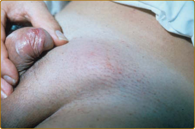
Figure 173-1 Lymphogranuloma venereum.
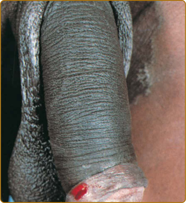
Figure 173-2 Lymphogranuloma venereum: soft painless erosion on the prepuce.
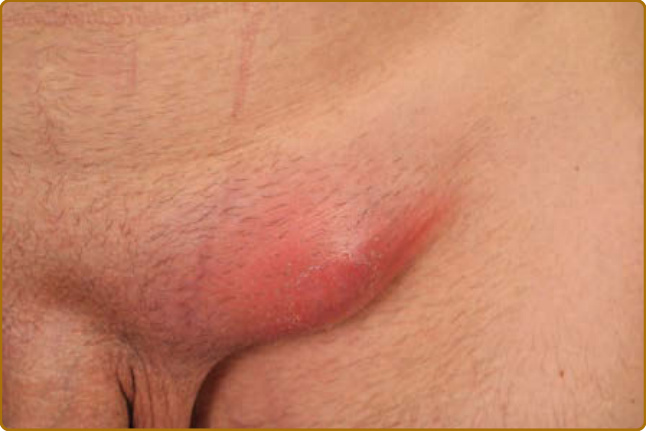
Figure 173-3 Lymphogranuloma venereum: lymph node involvement. Initially, the overlying skin is erythematous and indurated.
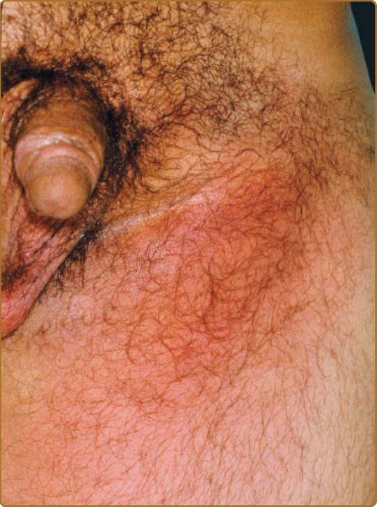
Figure 173-4 Early bubo consisting of unilateral enlargement and coalescence of inguinal lymph nodes. Note the absence of the primary lesion in this case. (Used with permission of Shukrallah Zaynoun, MD.)
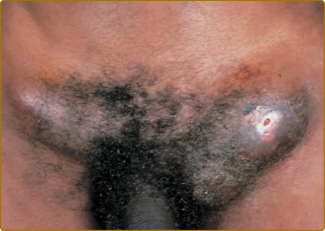
Figure 173-5 Lymphogranuloma venereum: bilateral, firm, immovable masses above the Poupart ligament.
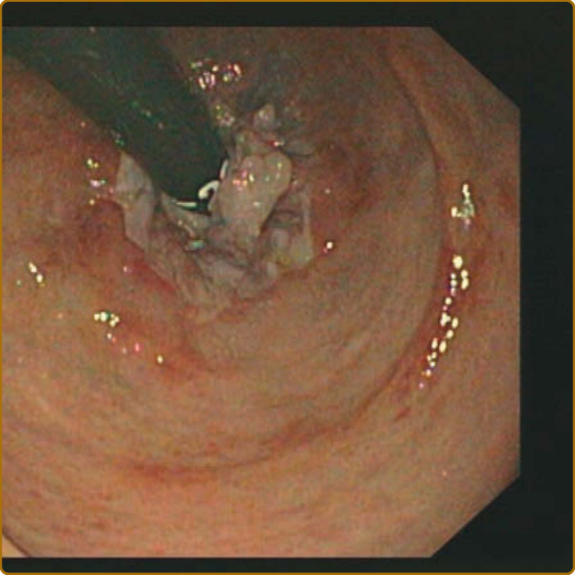
Figure 173-6 Lymphogranuloma venereum imitating Crohn disease.
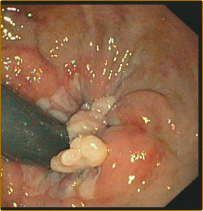
Figure 173-7 Exophytic tumor caused by lymphogranuloma venereum.
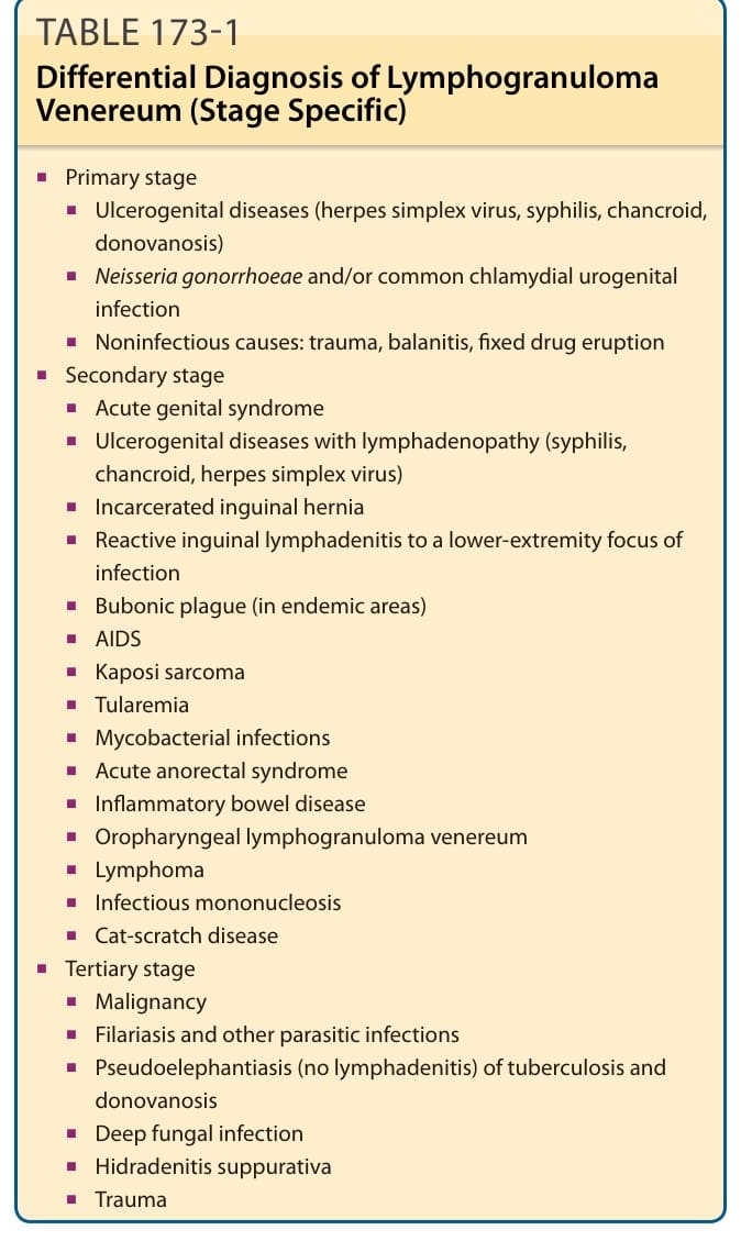
Table 173-1 outlines the differential diagnosis of LGV. In contrast to LGV primary stage, chancroid ulcers are usually larger and more painful, and donovanosis (granuloma inguinale) ulcers have abundant friable granulation tissue without associated lymphadenitis. Acute genital syndrome may be hard to differentiate from chancroid. Buboes containing little or no pus are, however, more likely to be caused by LGV. Suspecting LGV proctitis in HIV-positive MSM who present with signs and symptoms of Crohn disease (Fig. 173-6) or malignancy (Fig. 173-7) is important, even in the absence of LGV pathognomonic findings.68 Both conditions have similar proctoscopic findings; however, Crohn disease is more proximally localized. Studies from The Netherlands, the United Kingdom, and
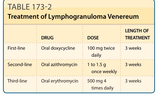
TABLE 173-2 Treatment of Lymphogranuloma Venereum