Melanoma
20
AT-A-GLANCE
■ Rising incidence worldwide in countries with white inhabitants, with highest incidence rates in Australia (35 new cases per year per 100,000 population), followed by North America (21.8 new cases per 100,000 population) and Europe (13.5 new cases per 100,000 population).
■ Risk factors include history of sunburns and/or heavy sun exposure, blue or green eyes, blonde or red hair, fair complexion, >100 typical nevi, any atypical nevi, prior personal or family history of melanoma, or p16 mutation.
■ Mean age of diagnosis is 63 years, with 15% being younger than 45 years.
■ Most common location is the back for men, and lower extremities followed by trunk for women, but can occur anywhere on the skin surface.
■ Features used for melanoma recognition: A (asymmetry), B (border), C (color), D (diameter, >5 mm in most common use), and E (evolution).
■ Follows a highly variable course and represents a heterogeneous disorder; surgically curable if diagnosed and treated in early phase, but potentially lethal with increased risk when diagnosed and treated late.
■ In the last decades, completely new and effective treatment options for metastatic melanoma approved with immunotherapies such as the immune checkpoint blockade (anti-CTLA4, anti-PD-1 antibodies) and targeted therapies like BRAF/MEK inhibitors leading to a median overall survival of 2 years in stage IV melanoma and the chance for a long-term tumor control.
INTRODUCTION
DEFINITIONS
DEFINITIONS
Melanoma (a word derived from the greek melas [dark] and oma [tumor]) is a malignant tumor arising from melanocytic cells and hence can occur anywhere where these cells are found. The most frequent type is cutaneous melanoma but melanomas develop also at the mucosal, the uveal, or even the meningeal membrane. Ten percent of melanomas are detected by lymph node metastases with so-called “unknown primary” and are likely to develop in the lymph node from preexisting nodal nevi.1
HISTORICAL PERSPECTIVE
HISTORICAL PERSPECTIVE
Cancer is a disease that probably accompanied human life from the very beginning. The earliest physical evidence of melanoma comes from diffuse melanotic metastases found in skeletons of Pre-Colombian mummies from Chancay and Chingas in Peru, dated to be about 2400 years old (radiocarbon). However, the first descriptions of the disease can be found in the writings of Hippocrates of Cos in the 5th century BC.2
Until a few years ago, the only cure was by surgery in the early stages. The first documented operation of a melanoma was done by the Scottish surgeon John Hunter in 1787. The preserved tumor is still housed in the Hunterian Museum at Lincolns Inn Fields in London.2 Until recently, in the unresectable metastatic setting, no treatment was available that could prolong patients’ survival. This was revolutionized in the last decade by the approval of immunotherapies, the so-called immune checkpoint blockers, and targeted treatments to the frequently found BRAF mutations.
EPIDEMIOLOGY
The incidence of melanoma has increased significantly worldwide over the last several decades. It is mainly a tumor of people with fair skin from more developed regions. There is a low annual incidence in the whole world population of 3.0 new cases per 100,000 population—thus, it is not one of the top 10 cancers worldwide.3 The highest incidence of melanoma is reported for Australia/New Zealand with about 35 new cases per year and 100,000 population, followed by Northern Europe and Northern America (Fig. 116-1). In the year 2016, an estimated 76,380 new cases of melanoma will be diagnosed in the United States of America translating to an annual incidence of 21.8 new cases per 100,000 population.4 In Europe, incidence rates are lower, with an annual incidence of 13.5 new cases per 100,000 population and large differences between European countries, with the highest incidence in Switzerland for men and in Denmark for women.5 Central and Eastern European countries have the lowest reported incidence rates in Europe. In the United States, incidence rises about 2.6% per year, leading to an increase of 33% for men and 23% for women between 2002 and 2006.6 Across all cancers, melanoma incidence is the most rapidly increasing in men and is only second to lung cancer in women. Invasive melanoma of the skin is the fourth most frequent site for cancer to occur in men and the sixth most frequent site in women.
20
Estimated cancer incidence for melanoma world map
Incidence ASR
Both sexes
Melanoma of skin
4.2+
1.6-4.2
0.89-1.6
0.48-0.89
<0.48
No Data
Source: GLOBOCAN 2012 (IARC)
By 2015, it is estimated that 1 in 50 Americans will develop melanoma in their lifetime.6
The median age for a melanoma diagnosis is 63 years with 15% being younger than 45 years. Incidence rises with age to a maximum between 55 and 74 years. Mortality data parallel incidence data, with a median age at death of 69 years.4 Even though melanoma accounts for only 4% of all skin cancer diagnoses each year in the United States, it is responsible for 75% of skin cancer deaths. Currently, one US citizen dies from a melanoma every hour. However, even though melanoma incidence is rising rapidly with a tripling from 1980 to 2003, it is not associated with a corresponding increase in melanoma deaths. This is probably based on earlier detection and hence better prognosis of the melanoma.6
CLINICAL FEATURES
CUTANEOUS FINDINGS
CUTANEOUS FINDINGS
The different subtypes of cutaneous melanoma can be distinguished clinically. However, these subtypes are not of prognostic significance itself, for example, a nodular or amelanotic melanoma might have a poorer prognosis compared to a superficial spreading melanoma but this is most likely based on a higher tumor thickness because of a later diagnosis.7 However, clinical heterogeneity of melanomas might be explained by genetically distinct types of melanoma with different susceptibility to ultraviolet light.8
SUPERFICIAL SPREADING MELANOMA (SSM)
SSM is the most common subtype, accounting for approximately 70% of all cutaneous melanomas. It is diagnosed most commonly on intermittently sunexposed areas, most frequently the lower extremity of women, and the upper back of men. Its classic clinical appearance best fits into the ABCD criteria (see section “Making a Diagnosis”), with irregular borders and irregular pigmentation, but it may present subtly as a discrete focal area of darkening within a preexisting nevus. The range of appearance of SSM is broad (Figs. 116-2A,B, and Fig. 116-3). Although varying shades of brown typify most melanocytic lesions, striking aspects of dark brown to black, blue-gray, red, and gray-white (which may represent regression) may be found in melanoma. SSM is the subtype of melanoma most commonly associated with preexisting nevi. The history of SSM is often of a lesion slowly changing over months to years. It may be mistaken for an atypical nevus or seborrheic keratosis.
NODULAR MELANOMA (NM)
NM is the second most common melanoma subtype and accounts for approximately 15% to 30% of all melanomas. The trunk is the most common site. NM is remarkable for rapid evolution, often arising over several weeks to months. NM more often lacks an apparent radial growth phase. It is more common
1983
20
A
C
E
B
D
F
1984
G
for NM to begin de novo than to arise in a preexisting nevus. NM typically appears as a uniformly dark blue-black or bluish-red raised lesion, but 5% are amelanotic (Fig. 116-2C,D). Early lesions often lack asymmetry, have regular borders, and are a uniform color. Amelanotic lesions may be mistaken for basal cell carcinoma, pyogenic granuloma, or hemangioma, whereas pigmented lesions may be mistaken for blue nevi or pigmented basal cell carcinomas. SSM and NM have the highest rate of mutations in the BRAF gene, with up to 56% of the melanomas harboring this alteration.8 There is also an association with multiple melanocytic nevi.
20
H
LENTIGO MALIGNA (LM) AND LENTIGO MALIGNA MELANOMA (LMM)
LM is a melanoma in situ with a prolonged radial growth phase that eventually becomes invasive and is then called LMM. LMM constitutes 10% to 15% of cutaneous melanomas. LM and LMM are diagnosed most commonly in the seventh to eighth decades in an older population than other types of melanoma—uncommon before the age 40. The most common location is on the chronically sun-exposed face, on the cheeks and
A H&E Melan-a
B H&E Melan-a
1985
20
nose in particular; the neck, scalp, and ears in men. Its pathogenesis is thought to be related to cumulative sun exposure rather than intermittent exposure. Clinical appearance is an initially flat, slowly enlarging, brown, frecklelike macule with irregular shape and differing shades of brown and tan, usually arising in a background of photodamage (Fig. 116-2E). Both LM and LMM often have clinically ill-defined borders, which may be obscured by background actinic damage consisting of lentigines, pigmented actinic keratoses, or ephelides. LM and LMM are associated with significantly higher rates of extensive subclinical lateral growth, resulting in higher recurrence rates with standard recommended margins and failure to completely excise the lesion. LM and LMM have the least common association with nevi, at 3% of cases, but the highest rate of association with desmoplastic melanoma (DM, see below). Molecularly, melanomas occurring in sundamaged skin carry c-KIT aberrations (mutations and copy number changes) more frequently than BRAF mutations, with up to 28% versus 6%.9
ACRAL LENTIGINOUS MELANOMA (ALM)
ALM is a subtype of melanoma with distinct differences in frequencies seen between ethnic groups. ALM constitutes only 2% to 8% of melanomas in whites but represents the most common form in darker-pigmented individuals (60% to 72% in African Americans and 29% to 46% in Asians). Although the proportion of ALM seen in darker-pigmented individuals is higher, the incidence of ALM is similar between whites and other ethnicities. ALM is diagnosed more often in an older population, with the median age of onset of 65. The most common site for ALM is the sole, with the palm and subungual locations following (Fig. 116-2F). The clinical appearance of ALM is mainly brown to black, but red with variegations in color and irregular borders can occur. Often ALM are misdiagnosed first as a plantar wart or hematoma, leading to a more advanced lesion upon diagnosis associated with poorer outcomes. ALM is not thought to be associated with sun exposure. Subungual melanoma, considered a variant of ALM, generally arises from the nail matrix, most commonly on the great toe or thumb (Fig. 116-2G). It appears as a brown to black discoloration or growth in the nail bed (Table 116-1). A widening, dark, or irregularly pigmented longitudinal nail streak (melanonychia striata) with or without nail dystrophy and nail plate elevation may be seen. Hutchinson sign, the finding of pigmentation on the proximal nail fold, may be noted with subungual melanoma. Benign lesions that mimic subungual melanoma include benign longitudinal melanonychia, subungual hematoma, pyogenic granuloma, or even onychomycosis with pigmentation or hemorrhage. In acral melanoma, the most frequent targetable mutation is the BRAF mutation (21%), followed by a c-KIT mutation (13%)10—c-KIT aberrations including
1986
■Lesion is isolated on a single digit
■Lesion is isolated on a single digit
■Occurrence in the fourth to sixth decade
■Occurrence in the fourth to sixth decade
■Abrupt pigmentation in a previously normal nail plate
■Abrupt pigmentation in a previously normal nail plate
■Pigmentation appears darker, larger, or blurred towards the nail matrix (proximally)
■Pigmentation appears darker, larger, or blurred towards the nail
matrix (proximally)
■Acquired pigmentation of the thumb, index finger, or large toe
■Acquired pigmentation of the thumb, index finger, or large toe
■Pigmentation occurring after a history of trauma, after ruling out subungual hematoma
■Pigmentation occurring after a history of trauma, after ruling out
subungual hematoma
■Individual with a history of melanoma
■Individual with a history of melanoma
■Pigmentation associated with nail dystrophy, or absence of nail plate
■Pigmentation associated with nail dystrophy, or absence of nail
plate
■Pigmentation extending to the periungual skin (Hutchinson sign)
■Pigmentation extending to the periungual skin (Hutchinson sign)
Braun R, Baran R, Frederique A, et al. Diagnosis and management of nail pigmentations. J Am Acad Dermatol. 2007;56(5):835-847.
copy number changes are higher, with up to 36% but with questionable clinical significance.9
DESMOPLASTIC MELANOMA (DM)
DM most commonly develops in the sixth or seventh decade on sun-exposed head and neck regions. The lesions typically have a firm, sclerotic, or indurated quality, and one-half are amelanotic (Fig. 116-2H). Approximately half of the lesions arise in association with the LM histologic subtype. Although often deeply invasive at the time of diagnosis with a tendency to perineural growth, DM is associated with higher local recurrence but lower nodal metastatic rates than other subtypes of melanoma when matched for depth of invasion. Results from a small study that involved 10 samples that investigated gene expression profiling demonstrated a molecular distinction between DM and nondesmoplastic melanoma.11 DMs reveal a high mutation burden most likely induced by UV radiation. BRAF and NRAS mutations are not found; instead, other genetic alteration known to activate the MAPK signaling cascade were identified, for example, mutations in neurofibromin (NF1) in more than 90% of cases.12,13
MUCOSAL MELANOMA
Melanoma can infrequently (1.3% of melanomas) arise on mucosal surfaces on the head and neck (conjunctival, intranasal, sinus, and oral cavities), genital, anorectal, or even urethral mucosa (Fig. 116-4).14 With the exception of the conjunctiva, patients present most often with delayed detection and a deeply pigmented, irregular lesion, often tumorous with signs of bleeding. As most of these lesions present initially with a radial growth phase manifesting a macular pigmentation, any suspicious area in these sites should be biopsied. Lesions of the conjunctiva are visible and appear increased in patients with atypical nevi.15 Mucosal melanomas are more frequent in women, especially
A
C
E
20
B
D
F
based on the genital tract melanomas. Melanomas on vulva and vagina account for about 50% of the mucosal melanomas among women. In both genders, the nasal cavity was otherwise the most frequent location for
mucosal melanomas. Anorectal melanomas account for only 16.5% of mucosal melanomas.14
Mucosal melanoma differs from cutaneous melanoma on the molecular level. Besides NF1, the most
1987
20
frequently occurring driver mutations are RAS alterations, consisting of NRAS and KRAS mutations with a frequency between 5% and 30% that lead to an activation of the MAPK pathway similar to BRAF mutations. Less than 10% of mucosal melanomas harbor BRAF mutations.16-18 Only in mucosal melanomas of vulvovaginal origin, the mutation frequency is higher, with up to 26% for BRAF and lower for RAS.19 More frequent than in cutaneous melanoma, mutations of the receptor tyrosine kinase c-kit can be found with varying frequencies in between 5% and 20% of mucosal melanomas.9,16,17 Here, frequency again seems to be higher in tumors of vulvovaginal or anorectal origin, compared with other sites.19,20 This mutation is targetable with multi-kinase inhibitors such as imatinib21; however, the mutation status of the tumors showed no association with patients’ survival outcomes.17
NEVOID MELANOMA
Nevoid melanoma describes a heterogeneous group of rare lesions that histologically resemble benign nevi by their symmetry and apparent maturation with descent in the dermis, thus with greater potential for misdiagnosis. Clues to their histologic diagnosis include marked hyperchromasia of the nuclei of the tumor cells, the presence of mitoses, and expansive growth of the dermal cells. Clinically, this may correspond to a tan papule or nodule, more often >1 cm in diameter on a young adult.22
SPITZOID MELANOMA
Spitzoid melanoma is a subtype of melanoma that clinically and histologically resembles a Spitz nevus, but tends to be larger and have asymmetry and irregular coloration. Features that favor the diagnosis of a Spitzoid melanoma over a benign Spitz nevus are large size (greater than 1 cm in greatest dimension); lesions with a thick invasive component (>2 mm Breslow thickness); lesions with numerous mitoses (especially any atypical forms), many cytologically atypical cells, and lesions that have a clinically concerning course such as very rapid growth in size or satellitosis. Clinical and histologic distinction between the two is often extremely difficult, and tumors with overlapping features of Spitz nevi and melanoma are classified as atypical Spitz tumors of uncertain biologic behavior (AST).23
Whereas common acquired nevi and melanoma often harbor BRAF mutations, NRAS mutations, or inactivation of NF1, Spitz tumors show HRAS mutations, inactivation of BAP1, or genomic rearrangements involving kinases like ALK, ROS1 and others. Additional genomic aberrations may abrogate various tumor-suppressive mechanisms and lead to atypia and malignant transformation. Comparative genomic hybridization (CGH) is generally able to differentiate Spitz tumors of benign or malignant behavior and may help classify the histologically ambiguous tumors, that is, the AST.24
1988
OTHER RARE TYPES
Several other rare and unusual types of melanoma are beyond the scope of this chapter, but previously reported in a comprehensive review, which include metaplastic melanoma, balloon cell melanoma, signetring cell melanoma, myxoid melanoma, rhabdoid melanoma, animal-type melanoma, and malignant blue nevus.22,25
NONCUTANEOUS FINDINGS
NONCUTANEOUS FINDINGS
Although most melanomas develop on the skin, there are rarer types of noncutaneous melanomas arising from extracutaneous melanocytes, for example, in the choroidea. In addition, mucosal melanomas (see before) are defined as noncutaneous melanomas as they can occur at mucosa of the ear, nose, and throat (ENT) region, intestine, or urinary tract. Melanomas of unknown primary that most often are diagnosed with lymph node metastases might develop from nodal nevi.1 As nodal nevi only occur in skin draining lymph nodes, the precursor lesion here might be nevus cells that escaped from melanocytic nevi of the skin.26,27
UVEAL MELANOMAS
Uveal melanomas account for about 5% of all melanomas and develop mainly in the choroid, followed by ciliary body and iris of the eye.28 Nevertheless, uveal melanoma is the most common primary intraocular malignancy. The median age at diagnosis is 58 years. Risk factors include the presence of a choroidal nevus, a nevus of Ota, and dysplastic nevus syndrome. There is an 8 times higher incidence rate in whites as compared to the black population, leading to the hypothesis that UV exposure increases the risk for uveal melanoma, but this has not been definitely proven.14
Clinically most patients present with painless loss or distortion of vision or the tumor is diagnosed in asymptomatic patients in routine ophthalmic screening. The risk of metastases could be linked to chromosomal aberrations. Patients with monosomy 3 (about 50% of patients) had a much poorer clinical outcome, with a disease-specific mortality of 75.1% for patients with monosomy 3 and 13.2% for patients with disomy 3 after 5 years, respectively.29 Patients usually die from liver metastases, the primary metastatic site in uveal melanoma probably based on the microenvironment in the liver that seems to support growth of the metastases there, for example, by secretion of the hepatocyte growth factor (HGF).30
COMPLICATIONS
COMPLICATIONS
If complications occur with melanoma, they are usually based on metastatic disease within organs leading to symptoms associated with the affected organ.
These include pain (any metastases), convulsion (brain metastases), instabilities (bone metastases), etc and later on all the symptoms associated with progression of the disease and death in the palliative setting. Relatively frequent cutaneous changes associated with melanoma are localized or diffuse hypo- or hyperpigmentation. The development of a melanoma-associated vitiligo as an accompanying autoimmune disease against melanocytes is reported to occur in up to 4% of patients and is associated with a better prognosis.31
Vitiligo has become especially frequent in patients treated with immune checkpoint blockers, and is also correlated with a better treatment response.32 In highly advanced patients, a diffuse cutaneous melanosis can occur that is characterized by a slate-grey pigmentation over the entire skin surface, mucosal membranes, and internal organs, with accentuation in photo-exposed areas. Such cases are often associated with melanuria, a darkening of the urine. Histopathologically, melanin deposits in the connective tissue and within macrophages in a perivascular distribution can be found. These most likely come from ischemic metastases distributed through the body via the blood.31
Rare complications based on paraneoplastic mechanisms described like dermatomyositis (Chap. 62)31 and autoimmune retinopathies also occur.33 Melanomaassociated retinopathies (MAR) present after the melanoma has been diagnosed as metastatic and are more frequent in men. Patients may develop vision problems years later, leading to latency from melanoma diagnosis to recognition of MAR on average after 3.6 years. Pathophysiologically, antiretinal antibodies against different structures like the bipolar cells, transducing, rhodopsin, and others can be found.
ETIOLOGY AND PATHOGENESIS
There are several risk factors for melanoma, with sun exposure and genetics being the 2 most important ones. Risk prediction models reveal in detail that the number of nevi, presence of freckles, history of sunburn, hair color, and skin color are the best measures to estimate melanoma risk.34,35 All these factors are influenced by the genetic background leading to a certain photosensitivity.
SUN EXPOSURE
SUN EXPOSURE
There is clear convincing evidence that sun exposure, and more specifically ultraviolet (UV) exposure, is a major environmental cause of melanoma, especially in high-risk populations. However, certainly not all melanomas are sun related. Bastian and others suggested a molecular classification for melanoma based on the hypothesis that clinical heterogeneity is explained by genetically distinct types of melanoma with different susceptibility to ultraviolet light.8
20
Epidemiologic studies suggest that periodic, intense sun exposure (particularly during the critical time period of childhood and adolescence) rather than long, continued, heavy sun exposure is most important in melanoma causation, termed the intermittent exposure hypothesis. Sunburn history, notably blistering and peeling burns, serves as a surrogate measure of intermittent intense sun exposure. In most melanoma risk prediction models, the history of sunburns is an important risk factor, not just in childhood,34-36
that is, the more sunburns in a lifetime, the higher the melanoma risk. One blistering sunburn in childhood more than doubles a person’s chances of developing melanoma later in life.37
The anatomic distribution of melanoma by body site demonstrates that intermittently exposed skin areas have the highest rates of developing melanoma. In men, the trunk, particularly the upper back, is the most common site for melanoma. In women, the lower legs, followed by the trunk, are the most common sites.38 These intermittently exposed areas are the most common areas to develop melanoma in younger persons. In older persons, there is a greater incidence of melanomas located on chronically exposed areas with maximal cumulative sun exposure. The face is the most common location for melanoma in older persons, with the addition of the neck, scalp, and ears as well, in older men.39
Several forms of artificial light have been associated with the development of melanoma, particularly psoralen and UVA light (PUVA) and UVB as well as tanning booths. The so-called PUVA Followup Study demonstrated increased rates of melanoma after PUVA exposure, with an incidence rate ratio of 9.3 approximately 20 years after PUVA therapy; these rates increase over time and are higher in patients exposed to high cumulative doses of PUVA.40 However, in 2 European cohorts of PUVA-treated patients, there was no increased risk for melanoma detected in contrast to nonmelanoma skin cancers such as squamous and basal cell carcinomas.41 There is also rising concern over tanning beds and melanoma risk, especially because exposure to the artificial UV radiation is intermittent in nature. Any exposure to artificial tanning devices significantly increases the risk of cutaneous melanoma, and longer duration of bed use, younger age at first exposure, and higher frequency of use are associated with a significantly elevated risk (odds ratio 1.69).42 Moreover, indoor tanning also has been associated with accelerated skin aging and ocular melanoma.43
Pathophysiologically, DNA-damaging, carcinogenic, inflammatory, and immunosuppressive properties of UV radiation contribute to initiation, progression, and metastasis of primary melanoma.37,44 UV-A (320 to 400 nm) and UV-B (280 to 320 nm) have a significant impact, with UV-B directly damaging the DNA and UV-A mainly by the production of reactive oxygen species (ROS). The final proof of the carcinogenic property of UV on melanoma development was given by a large Australian clinical trial. In 1992, a total of 1621 randomly selected residents from Queensland between
1989
20
25 and 75 years of age were randomly assigned to daily or discretionary sunscreen use to head and arms until
2006. Ten years after trial cessation, the rate of primary melanomas was halved in the daily sunscreen group, with 11 new primary melanomas instead of 22 in the discretionary sunscreen group.45
SKIN PHENOTYPE
SKIN PHENOTYPE
Light skin pigmentation, blond or red hair, blue or green eyes, prominent freckling tendency, and tendency to sunburn with Fitzpatrick skin phototype I–II are phenotypic features associated with an increased risk of melanoma of 2- to 3-fold.34,46 Melanoma occurs much less frequently in Type V–VI skin, suggesting that skin pigment plays a protective role. This is explained of course by different photosensitivity and ability to tan.
MELANOCYTIC NEVI
MELANOCYTIC NEVI
There is an increased risk of melanoma associated with nevi, both in a quantitative (ie, number of nevi) and qualitative (ie, typical vs atypical nevi) manner.34,47
Adults with more than 100 clinically typical-appearing nevi, children with more than 50 typical-appearing nevi, and any patient with atypical nevi are at risk. The presence of a solitary dysplastic nevus doubles the risk of melanoma, while having 10 or more atypical nevi is associated with a 12-fold elevation of risk.48 Nevi more often serve as a genetic marker of increased risk rather than a premalignant lesion, as most melanomas arise de novo. In a study of 1606 patients with melanoma, only 26% of the melanomas were histologically associated with nevi (43% of these atypical nevi, 57% other nevi).49 However, sun exposure plays an important role to acquire melanocytic nevi, particularly in early childhood. Hence, the amount of melanocytic nevi as a risk factor for melanoma development is influenced by genetic as well as environmental factors.47
There is a recognized risk of melanoma development in large congenital nevi, which varies depending on the size of the lesion.50,51 Many series define large congenital nevi as greater than 20 cm in diameter in adulthood, and lifetime risks for developing melanoma are generally accepted to be in the 2% to 10% range. In a large review of more than 2500 patients with a large congenital melanocytic nevus, 2% developed a melanoma at a median age of 12.6 years (range birth to 58 years). In 74% of patients, the melanocytic nevus was greater than 40 cm in diameter, and 94% had satellite nevi.51 Patients with large congenital nevi located on the posterior axis (paraspinal, head, and neck regions) or in conjunction with multiple satellite lesions are at risk for neurocutaneous melanosis, with an increased risk of developing melanoma in the CNS. Melanomas developing on large congenital nevi are usually detected late and hence have a bad prognosis, with more than 50% of them being fatal.51 For small- to
1990
CDKN2A p16 CDK4 POT1 TERT BAP1 BRAF NRAS HRAS NF1 c-KIT
CDKN2A p16 CDK4 POT1 TERT BAP1 BRAF NRAS HRAS NF1 c-KIT
medium-sized congenital nevi, the melanoma risk is similar to any other area of skin.50 Thus, prophylactic excision of small- and medium-sized congenital nevi is usually unwarranted.
FAMILY HISTORY
FAMILY HISTORY
Patients with familial melanoma are estimated to account for approximately 5% to 12% of all patients with melanoma.46 Having one first-degree relative with melanoma doubles the risk of melanoma, whereas having 3 or more first-degree relatives with melanoma increases the risk 35- to 70-fold.52 Some of this risk may be attributed to shared risk factors such as skin phenotype, multiple nevi, and excessive sun exposure. The association between familial melanoma and multiple atypical nevi has historically been given various names, including B-K mole∗ syndrome, familial atypical multiple mole-melanoma syndrome, and dysplastic nevus syndrome. Patients with familial melanoma typically have earlier-onset melanoma and multiple primaries as well as atypical nevi. In addition, patients with familial melanoma have an increased risk of internal cancers, such as pancreatic cancer or CNS tumors.46
Different genes are responsible for the development of inherited melanoma (Table 116-2) and genetic alterations typically increase cancer risk via 3 major mechanisms: the activation of oncogenes, the loss of tumor suppressor genes, or increased chromosomal instability. Inherited mutations in CDKN2A, CDK4, POT1, and TERT confer a 60% to 90% lifetime risk of melanoma.
CDKN2A-CDK4-TP53 PATHWAY MUTATIONS
Germline mutations in the chromosome 9p21 tumor suppressor gene, cyclin-dependent kinase inhibitor 2A (CDKN2A), account for approximately 40% of hereditary melanoma cases (≥3 melanomas in one lineage). In countries with high levels of UV exposure, the lifetime melanoma risk in carriers of the CDKN2A mutation
∗B-K=initials of the first 2 described related patients.
is 76% (United States) and 91% (Australia), whereas the risk is lower in countries with low levels of UV exposure (58%, United Kingdom).46 Individuals with germline CDKN2A mutations also exhibit a higher risk of pancreatic cancer; an estimated 15% of individuals with a mutant allele will develop pancreatic cancer in their lifetime.53 Bona fide deleterious point mutations in CDKN2A are relatively uncommon in primary melanoma tumors although homozygous deletions of this gene may obscure the true rate of somatic loss in melanoma. CDKN2A encodes 2 gene products: p16 (also known as INK4a, inhibitor of kinase 4a) and p14ARF
(alternative reading frame). p16 is a cell-cycle regulator that binds and inhibits cyclin-dependent kinases Cdk4 or Cdk6, thereby inhibiting progression of cells through the G1 phase of the cell cycle (Fig. 116-5). If p16 function is absent or inactivated by mutation, unrestrained Cdk4 activity phosphorylates the retinoblastoma protein, thereby releasing the transcription factor E2-F and inducing S-phase entry. This sequence culminates in enhanced cellular proliferation, which, in the absence of checkpoint regulation, results in unrestrained growth and neoplasia. The binding partner of the p16 protein is Cdk4. Only a handful of families worldwide thus far have been reported to carry hereditary mutations in CDK4, while somatic mutations in this gene also have been detected in some melanoma cell lines. Functional studies suggest that mutations in Cdk4 render the cyclin-dependent protein kinase resistant to p16 inhibition, resulting in a phenotype identical to that from p16 loss.
20
The p14ARF protein from CDKN2A inhibits a cellular oncogene Hdm2, which in turn accelerates the destruction of the p53 tumor-suppressor gene.54 Complete loss of CDKN2A also leads to abrogation of p14ARF and loss of p53 function. Thus, this single locus can inactivate both the retinoblastoma protein and p53 pathways and probably explains the low rate of direct TP53 mutagenesis in melanoma.
GENES INVOLVED IN TELOMERE MAINTENANCE
POT1 and TERT mutations are prevalent in 9% of patients with hereditary melanoma and, when normally functioning, contribute to protection of exposed chromosomal ends.46 Increased TERT activity may allow unlimited cell division and subsequently promote cancer progression.
DIAGNOSIS
Early detection is the key to improving prognosis in melanoma, as the risk of metastases increases with infiltration depth of the primary. Although melanoma may have a characteristic appearance, there is no single clinical feature that ensures or excludes a diagnosis of melanoma. Change in color and increase in size (or a new lesion) are the 2 most common early characteristics noticed by patients that may be useful in discriminating between melanoma and other benign lesions.55
CDKN2A-CDK4-TP53 pathway
CDKN2A CDKN2A
P16INK4A
P27kip1 P21cip1 RB E2F
P P
RB
CDK4/6
cyclinD CDK2
cyclinE
e
s
a
h
P
Restriction point
1-
S
G
P
h
a
s
e
E2F
e
s
a
h
P
M
2
P
G
h
a
s
e
P14ARF
Hdm2
DNA damage
p53
1991
20
PHYSICAL EXAMINATION
PHYSICAL EXAMINATION
The skin examination should be conducted under optimal lighting and encompass the entire skin integument, including the scalp, external ocular/conjunctivae, oral mucosa, groin, buttocks, and palms/soles/web spaces. Melanomas in hidden anatomic sites are associated with thicker tumors at diagnosis, often as a result of later detection.56 Clinical diagnosis of melanoma can be made in about 80% to 90% of cases. The well-known ABCDE acronym for melanoma detection continues to be a useful tool for the lay public and physicians.57,58
A stands for asymmetry (one half is not identical to the other half), B for border (irregular, notched, scalloped, ragged, or poorly defined borders as opposed to smooth and straight edges), C for color (having varying shades from one area to another), D for diameter (ie, greater than 5 mm), and E for evolution (changes in the lesion over time). Lesions having these characteristics may potentially represent melanoma. Studies have found the sensitivity of the ABCDE checklist (Table 116-3) to be very high, but the specificity much lower.59
Another diagnostic aid that is useful in detecting melanoma is the “ugly duckling” sign.60 A pigmented lesion that is different from other pigmented lesions on a particular individual should be approached with a high index of suspicion. This is based on the premise that within an individual, nevi should globally share a common appearance or family resemblance. Even in an individual with multiple atypical nevi, the nevi should be morphologically similar.
DERMOSCOPY
DERMOSCOPY
Dermoscopy (also known as epiluminescence microscopy, dermatoscopy, incident light microscopy, and surface microscopy) is a noninvasive technique in which a handheld device is used to examine a lesion through a film of liquid (eg, immersion oil), using nonpolarized light (contact dermoscopy), or the lesion is examined under polarized light without a contact medium (noncontact dermoscopy). In experienced hands, it may improve both the sensitivity and specificity for the clinical diagnosis of melanoma and other pigmented and nonpigmented lesions.61,62 Morphologic features (Table 116-4) that are otherwise not visible to the naked
■A = Asymmetry
■B = Border
■C = Color
■D = Diameter
■E = Evolution
■E = Evolution
1992
aDoes not apply to nodular or desmoplastic melanoma.
■Atypical pigment network
■Atypical pigment network
■Negative network
■Negative network
■Atypical dots or globules
■Atypical dots or globules
■Irregular streaks
■Irregular streaks
■Regression structures
■Regression structures
■Blue-white veil
■Blue-white veil
■Shiny white lines
■Shiny white lines
■Atypical blotch
■Atypical blotch
■Polymorphous vessels
■Polymorphous vessels
Data from Wolner Z, Yelamos O, Liopyris K, et al. Enhancing skin cancer diagnosis with dermoscopy. Dermatol Clin. 2017;35(4):417-37.
eye are observed using this technique that allows visualization of microstructures of the epidermis, the dermoepidermal junction, and the papillary dermis (Fig.116-3). Different diagnostic algorithms using dermoscopic findings have been developed for melanoma, including the ABCD rule, the 7-point checklist, pattern analysis, Menzies method, modified ABC-point list, and CASH (color, architecture, symmetry, and homogeneity).63-66
Several studies have compared these methods to determine the most effective method for dermoscopic detection of melanoma.63,64 Pattern analysis, which provides an overall impression of multiple dermoscopic patterns without rigid rules, based primarily on a subjective, simultaneous evaluation of a number of different criteria, is the most widely used method among experienced users of dermoscopy for evaluating pigmented lesions.67
Digital dermoscopy or digital epiluminescent microscopy permits computerized digital dermoscopic images to be retrieved and examined at a later date so that comparisons can be made and changes detected over time. There are also a number of commercially available automated computer image analysis programs, devices that incorporate image analysis algorithms to digital dermoscopic images, providing objective measurement of changes over time. Sequential digital dermoscopy was shown to improve early diagnosis of melanoma compared to standard dermoscopy.68,69
The use of body or lesional photography can help to monitor minor changes in melanocytic lesions, particularly in patients with many nevi. Finally, confocal scanning laser microscopy and multispectral digital dermoscopy are among a number of new imaging techniques being evaluated for early detection of melanomas.70,71
HISTOLOGY
HISTOLOGY
Patients with lesions clinically suspicious for melanoma should, whenever possible, undergo prompt excisional biopsy with narrow margins. A wider margin should be avoided to permit the most accurate
subsequent SLNB if indicated.72 However, if the lesion is large and/or located on anatomic areas such as the palm/sole, digit, face, or ear, an incisional skin biopsy may be performed in the most elevated or darkest area of the lesion, with a strong appreciation that the clinically most suspicious area may not always correlate with the thickest portion of the lesion. There is no evidence that biopsy or incision of a primary melanoma leads to “seeding” of tissue and adversely affects survival.73 Excisional biopsy performed after incisional biopsy of melanoma with ≥50% of the lesion remaining resulted in significant upstaging in 21% of patients, and change in SLNB consideration in 10% of patients in one large study.74 Hence, a wider margin should be taken after complete excision with narrow margins and histologic evaluation of the whole lesion. Excisional biopsy, if possible, is important because symmetry of the whole lesion is one major criterion in differentiating a benign melanocytic nevus from melanoma. The histologic diagnosis of melanoma is based on the assessment of a constellation of findings, including both architectural and cytologic features. No single feature is diagnostic. The major architectural features of melanoma include asymmetry, poor
Superficial spreading melanoma
Penetrates basement membrane Spreads along epidermis
A
Nodular melanoma
Primarily deep invasion
B
Lentigo maligna melanoma
Site of invasion into dermis Multifocal atypical melanocytes
Malignant cells in dermis C
20
circumscription (cells at the edge of the lesion tend to be small, single, and scattered rather than nested), and large size (>5 to 6 mm). Nests of melanocytes in the lower epidermis and dermis tend to vary in size and shape, and to become confluent. There is a lack of maturation of nests with descent into the dermis. In addition, single melanocytes dominate over nests and show an aberrant distribution (Fig. 116-3).75 The presence of melanocytes above the basal layer (pagetoid spread), usually considered diagnostic of melanoma in situ, should be assessed cautiously in the context of other findings, as pagetoid spread may be seen in benign lesions including Spitz nevi and nevi in special anatomic regions (vulvar nevi, acral nevi). The different subtypes of melanoma may also have histopathologic differences (Fig. 116-6), especially DM, which is composed of strands of elongated, spindle-shaped cells that often infiltrate deeply in a sarcomatoid pattern. Immunohistochemistry may be useful for the diagnosis of melanoma, especially in poorly differentiated neoplasms with little or no pigment (ie, amelanotic melanomas), spindle cell tumors, or tumors with pagetoid spread that are not obviously melanoma. S100 and Sox10 proteins are expressed by almost all melanomas,
1993
20
but also by melanocytic nevi, and other tumor types, including cutaneous neural tumors. HMB-45 is a monoclonal antibody with high specificity for melanoma cells. Melan-A and MART-1 (melanoma antigen recognized by T cells) are the names given independently to the same gene encoding a melanocytic differentiation antigen expressed in the skin and retina. Melan-A is broadly expressed in benign and malignant melanocytic lesions. It is more sensitive than HMB-45 and more specific than S100 for melanoma. Search for the microphthalmiaassociated transcription factor (MiTF) may be useful, especially in amelanotic melanomas, as it is a marker in the nucleus, whereas all other markers are mainly intracytoplasmic.76 Immunohistochemically, DMs commonly express only S100 and Sox1077 and lack other melanocytic markers like HMB-45, Melan-A, and MiTF. In nevoid melanomas staining for the proliferation rate, for example, with Ki-67, might be helpful to uncover malignancy. In addition to diagnosis, histology gives further clinically important information on the infiltration depth (Breslow thickness) and ulceration status—features needed for AJCC (American Academy of Dermatology) classification and prognostic evaluation.78 Further diagnostic investigations are stage dependent as the risk to metastasize increases with tumor stage.78
LABORATORY TESTING
LABORATORY TESTING
As early as in 1954, increased levels of lactate dehydrogenase (LDH) were detected in the serum of melanoma patients.79 Ever since, the value of LDH as a tumor marker for melanoma has been discussed. S100B in the serum is a bit more specific than LDH but lacks sensitivity. For that reason, S100B can be detected and monitored in clinical followup as an additional marker to detect progression of the disease.80 Hence, blood investigations to measure for tumor markers play a minor role in the diagnosis of melanoma, but are mainly used to monitor the clinical course. Testing of the nonspecific tumor marker LDH is indicated only for patients with distant metastatic disease as it is needed for AJCC classification and prognostic evaluation.78,81
Other tumor markers like melanoma-inhibiting activity (MIA), tumor-associated antigen 90 immune complex (TA90IC), and YKL-40 are under investigation and not used in routine clinical practice. It is not clear as of this writing if they add a benefit to S100B and LDH.79
IMAGING
IMAGING
REGIONAL SKIN AND LYMPH NODE ULTRASOUND
Patients should be evaluated for regional spread by careful palpation of lymph nodes first, particularly
1994
the primary echelon nodal basin(s). The concepts of aberrant lymphatic drainage pathways to unexpected nodal basins and interval nodes located between the primary site and the expected regional nodal basin have taught physicians to search for clinically detectable nodal disease in unexpected locations.82 Skin and lymph node ultrasonography is perhaps the most sensitive noninvasive method to detect small nodal metastases and is much more sensitive than clinical examination.83 Lymph node metastases are characterized by a ballooned shape, loss of central echo, and peripheral perfusion.84 If a lymph node or a dermal/ subcutaneous nodule in the regional area of the primary is found, an excisional biopsy should be performed if possible.
TOMOGRAPHY
Routine imaging and hematologic tests in asymptomatic patients rarely identifies occult systemic disease.85 Tomographic investigations like computed tomography (CT), magnet resonance imaging (MRI) and positron emission tomography (PET) are generally not recommended at the stage of primary melanoma.86,87 The rate of false positive findings is far too high (8%-15%), for example, unspecific lung lesions,88 which leads to additional cost of followup studies and possibly invasive procedures, as well as increased patient anxiety. Unfortunately, there are no data demonstrating improved survival rates if metastases are detected when clinically asymptomatic versus early symptomatic stage IV disease. However, especially in high-risk melanomas (>4 mm Breslow thickness), the risk for hematologic spread increases, and tomographic imaging can be justified. Distant metastases are more likely in patients with regional metastases. However, in a retrospective analysis of 185 patients with a positive sentinel lymph node (SLN), only 1 patient (0.5%) had detectable metastatic disease on the imaging. Hence, imaging with CT, MRI, or PET is indicated only in high-risk and known metastatic melanoma and recommended to be performed as clinically indicated. This further emphasizes the importance of a thorough melanoma-focused review of systems and physical examination.
SENTINEL LYMPH NODE BIOPSY (SLNB)
SENTINEL LYMPH NODE
BIOPSY (SLNB)
The SLN is per definition the first draining lymph node in the lymphatic draining system of the primary tumor. SLNB is a powerful staging and prognostic tool which may be used to detect occult micrometastases in regional lymph nodes,89 and represents the best baseline staging test for detection of occult nodal metastasis in the subset of melanoma patients where it is indicated. SLNB is far more sensitive and accurate at detecting microscopic metastases than PET scan, CT scans, or ultrasonographic imaging combined with
lymph node fine-needle aspiration.85 In a meta-analysis of 34 studies, a Breslow thickness of <1 mm was associated with a positive SLN in 5.6% of patients.90 This rate was higher (19.5%) in patients younger than 40 years and a tumor with 0.75 to 1 mm thickness as well as in ulcerated primary melanomas and melanomas with detectable mitoses.91-93 For patients with lesions of a Breslow thickness between 1 and 4 mm Breslow, 25.2% had a positive SLN, whereas those with lesions >4 mm in thickness had a 50% likelihood of SLN positivity.92,94
Based on this risk stratification, SLNB is recommended in patients with a melanoma ≥1 mm Breslow thickness.89,90 In addition, it can be offered for patients with thinner melanomas (0.8-1 mm) or even <0.8 mm if additional risk factors such as high mitotic index or lymphovascular invasion exists, especially in the setting of young age.91
In current practice, technetium-99–labeled radiocolloid solution, often complemented with vital blue dye, allows for detection of the SLN >98% of the time in skilled hands.95 Ideally, the procedure should be performed at the same time as wide local excision (WLE) of the primary melanoma for greatest accuracy. SLNB may not be accurate if performed after WLE in areas of ambiguous or surgically altered lymphatic drainage or following a local skin flap due to radiocolloid and dye injection location away from the true primary site.96 Once removed, the SLN(s) must be assessed with serial sectioning, using standard hematoxylin and eosin (H&E) techniques often combined with immunohistochemical stains such as S100, HMB-45, and/or Melan-A. The use of immunostains increases sensitivity for melanoma and has resulted in the upstaging of up to 10% to 20% of patients.97 In melanoma, even single melanoma cells in the immunostains of the sentinel define the lymph node as sentinel positive (N1a). In 2006, Morton et al published the third interim analysis of the multicenter sentinel lymphadenectomy trial (MSLT)–I, the first prospective randomized controlled clinical trial of SLNB in melanoma.98 In this analysis, 1269 clinically node-negative patients with newly diagnosed melanoma 1.2 to 3.5 mm in depth were randomized to wide excision and nodal basin observation versus wide excision and SLNB. Patients with a positive SLN received an immediate complete lymph node dissection (CLND), and patients in the observation arm also received CLND if they developed regional lymph node metastasis. SLN status was demonstrated to be a powerful predictor of survival, as those with a negative SLN had a 5-year survival of 90.2%, versus 72.3% 5-year survival for those with a positive SLN. Recently, results of the DECOG-SLT trial have been published. In this German trial, almost 500 patients with a positive SLN with an at least 1-mm metastasis were randomized to obtain CLND or observation. This led to an improvement in local recurrence rate but did not lead to a survival benefit.99 Final analysis of both trials confirm these results with no significant benefit of an immediate CLND. Hence, this procedure is not recommended any more in patients with only microscopic nodal disease. However, Sentinel positive patients might be selected for adjuvant therapy.
20
Diagnostic algorithm
Clinical evaluation
• Medical history
• Physical examination
• Dermoscopy, computer-assisted diagnostics if necessary
Excisional biospy (at maximum 5-mm margins)
Staging
• Regional skin and lymph-node ultrasonography
• At Breslow index ≥4 mm consider tomographic imaging
Pos.
Neg.
Treatment according to melanoma stage SLNB At Breslow index ≥1 mm or below (if additional risk factors such as high mitotic index, lymphovascular invasion and younger age) consider SLNB
DIAGNOSTIC ALGORITHM
DIAGNOSTIC ALGORITHM
See Fig. 116-7.
DIFFERENTIAL DIAGNOSIS
See Table 116-5.
■Atypical nevus: ABCD criteria help to differentiate
■Atypical nevus: ABCD criteria help to differentiate
■Seborrheic keratosis: no pigment net in dermoscopy
■Seborrheic keratosis: no pigment net in dermoscopy
■Basal cell carcinoma, especially if pigmented: more black than brown, prominent vasculature
■Basal cell carcinoma, especially if pigmented: more black than
brown, prominent vasculature
■Hemangioma: can be difficult to distinguish from amelanotic melanoma
■Hemangioma: can be difficult to distinguish from amelanotic
melanoma
■Pyogenic granuloma: especially difficult to distinguish from amelanotic acral melanoma
■Pyogenic granuloma: especially difficult to distinguish from amela-
notic acral melanoma
■Solar lentigo, pigmented actinic keratosis, flat seborrheic keratosis: can mimic lentigo maligna melanoma
■Solar lentigo, pigmented actinic keratosis, flat seborrheic keratosis:
can mimic lentigo maligna melanoma
■Plantar wart: might be hard to distinguish from acral melanoma
■Plantar wart: might be hard to distinguish from acral melanoma
■Hematoma: especially acral or subungual hematoma
■Hematoma: especially acral or subungual hematoma
1995
■Longitudinal melanonychia: look for Hutchinson sign
■Longitudinal melanonychia: look for Hutchinson sign
20
CLINICAL COURSE AND PROGNOSIS
Melanoma can progress to different stages of the disease, starting with the primary (stage I/II), regional metastases (stage III) and distant metastases (stage IV) (Fig. 116-8). In general, prognosis and survival rates worsen with increasing stage. At initial presentation, approximately 85% of patients have localized disease, 10% have regional metastatic disease, and 5% have distant metastatic disease already.78
STAGES I AND II MELANOMA
STAGES I AND II
MELANOMA
In general, the 5- to 10-year survival for patients with localized thin primary melanoma <1 mm in Breslow depth is more than 90%. Melanoma typically recurs in a predictable fashion, first in a local and regional distribution, then to distant sites. It is also recognized that melanoma may bypass the regional nodes with direct hematogenous dissemination. The majority of recurrences manifest in the first 5 years after diagnosis and treatment, depending on tumor thickness and other prognostic features of the primary lesion. However, melanoma can recur at any time, and the incidence of late recurrence 10 or more years after initial diagnosis is approximately 1% to 5%.100
STAGE III MELANOMA
STAGE III MELANOMA
Stage III melanoma represents a broad range of patients with a diverse clinical outcome, from patients with microscopic nodal disease (IIIA) to patients with bulky clinical nodes or in-transit metastases (IIIC) (Fig. 116-8). The general overall 5-year survival range of 38% to 78% is wide, primarily related to several variables such as the number of positive lymph nodes (most important); tumor burden within a lymph node (microscopic vs macroscopic); age; and ulceration status as well as Breslow thickness of the primary melanoma (Table 116-6).78 There is a significant decline in survival in melanoma patients who present with clinically evident macroscopic nodal disease versus those with microscopic nodal disease identified via SLNB.
STAGE IV MELANOMA
STAGE IV MELANOMA
Melanoma is known for its propensity to metastasize to virtually any organ and also for its highly variable clinical course. Melanoma metastasizes to nonvisceral sites: distant skin/subcutaneous tissue and distant lymph nodes in approximately half of the stage IV cases (42% to 57%) (Fig. 116-8). The most
1996
A
B
C
T CLASSIFICATION THICKNESS (mm) ULCERATION STATUS
T1 ≤1.0 a: <0.8 mm without ulceration b: <0.8 mm with ulceration or 0.8-1 mm (with or without ulceration)
T2 1.01-2.0 a: without ulceration b: with ulceration
T3 2.01-4.0 a: without ulceration b: with ulceration
T4 >4.0 a: without ulceration b: with ulceration
N CLASSIFICATION NUMBER OF METASTATIC NODES NODAL METASTATIC MASS
n1
1
a: clinically occult b: clinically apparent c: pure in-transit met(s)/satellite(s) without metastatic nodes
N2 2-3 a: only occult metastases b: at least one clinically apparent metastasis c: in-transit met(s)/ satellite(s) with 1 metastatic node (clinically occult or apparent)
N3 ≥4 a: only clinically occult metastases b: at least one clinically apparent metastasis c: in-transit met(s)/ satellite(s) with at least 2 metastatic nodes (clinically occult or apparent)
M CLASSIFICATION SITE SERUM LACTATE DEHYDROGENASE
m1a Distant skin or soft tissue including muscles, or nodal metastases
M1a(0): Normal M1a(1): Elevated
M1b Lung metastases (with or without M1a involvement)
M1b(0): Normal M1b(1): Elevated
M1c All other visceral metastases without CNS metastases (with or without M1a or M1b involvement)
M1c(0): Normal M1c(1): Elevated
M1d CNS metastasis
M1d(0): Normal M1d(1): Elevated
M1d CNS metastasis (with or without M1a, M1b, or M1c involvement
M1d(0): Normal M1d(1): Elevated
(with or without M1a, M1b, or M1c involvement
20
common visceral sites are the lungs (18%-36%), liver (14%-20%), brain (12%-20%), bone (11%-17%), and GI tract (1%-7%). Once metastases to distant sites have been detected, median survival without any treatment is approximately 6 to 9 months. With the help of targeted and immunotherapies this can be increased to 2 years, and even long-term tumor control can be achieved.101,102
METASTATIC MELANOMA OF UNKNOWN PRIMARY (MUP)
METASTATIC MELANOMA
OF UNKNOWN
PRIMARY (MUP)
Metastatic melanoma of unknown primary (MUP) is defined as the presence of histologically confirmed melanoma in a lymph node, visceral site, or distant skin/subcutaneous tissues without a history or evidence of a primary cutaneous, mucosal, or ocular melanoma. This situation occurs in 2% to 5% of all cases of melanoma. Approximately 60% of these involve the lymph nodes and might have developed from nodal nevi.1 The remaining involve distant sites, typically skin/subcutaneous tissue, and less commonly lung, brain, or GI tract. MUP has similar survival rates compared to equivalently staged cutaneous melanomas of known primary origin. MUP metastatic to lymph nodes had 5- and 10-year survival rates of 46% and 41%, which is comparable to the equivalent stage III melanoma of known primary.103 Melanoma of unknown primary metastatic to viscera had a median survival of 6 months.104
Evaluation of melanoma of unknown primary should involve a complete skin examination looking for a primary site. Wood lamp may be used to identify subclinical hypopigmented areas, which may suggest a regressed primary lesion. In melanoma of unknown primary metastatic to a lymph node, particular attention should be focused on the skin areas that drain to the nodal basin involved. A complete history of previously excised skin lesions should be reviewed. The patient should be referred for proctoscopic, gynecologic, ocular, and nasal mucosal examinations, when warranted. However, only rarely a primary tumor is detectable, and hence more invasive procedures like gastroscopy/colonoscopy should be avoided without any clinical hints for a melanoma at these locations. Staging workup and treatment should be performed as for the equivalent clinical stage of known primary.
THE AJCC CLASSIFICATION
THE AJCC CLASSIFICATION
Accurate staging forms the basis for prognosis and treatment, which is invaluable for the majority of patients in their informed decision-making process. The American Joint Committee on Cancer (AJCC) published a revised staging system for melanoma in 2017
1997
20
CLINICAL STAGINGa PATHOLOGIC STAGINGb
T N M T N M
0 Tis N0 M0 Tis N0 M0
IA T1a N0 M0 T1a/b N0 M0
IB
T1b T2a N0 N0 M0 M0 T2a
N0
M0
IIA
T2b T3a N0 N0 M0 M0 T2b T3a N0 N0 M0 M0
IIB
T3b T4a N0 N0 M0 M0 T3b T4a N0 N0 M0 M0
IIC T4b N0 M0 T4b N0 M0
Any T
N1 N2 N3
IIIc
M0
IIIA
T1a/b–
N1a or N2a N1a or N2a
M0
T2a
M0
IIIB
T0 T1a/ b–T2a T2b/T3a
N1b/c N1b/c or N2b N1a-N2b
M0 M0
M0
IIIC
T0 N2b/c or N3b/c M0
T1a–T3a N2c or N3a/b/c M0
T3b/T4a T4b Any N N1a-N2c M0 M0
IIID
T4b N3a/b/c M0
IV Any T Any N Any
Any T Any N Any
IV Any T Any N Any M1 Any T Any N Any M1
M1
M1
aClinical staging includes microstaging of the primary melanoma and clinical/radiologic evaluation for metastases. By convention, it should be used after complete excision of the primary melanoma with clinical assessment for regional and distant metastases.
bPathologic staging includes microstaging of the primary melanoma and pathologic information about the regional lymph nodes after partial or complete lymphadenectomy. Pathologic stage 0 or stage IA patients are the exception; they do not require pathologic evaluation of their lymph nodes.
cThere are no stage III subgroups for clinical staging. M, metastasis classification; N, node status; T, tumor size. From Amin MB et al. AJCC Cancer Staging Manual, 8th ed. 2016.
(Tables 116-6 and 116-7).78,105,106 The 2017 melanoma staging system was obtained from a data set of >46,000 melanoma patients from 10 centers worldwide. This represents the largest body of AJCC melanoma data analyzed in an evidence-based approach and incorporates many patients more accurately staged using SLNB. The tumor size, node status, metastasis classification (TNM) system continues to form the backbone of the staging system, in which T describes the extent or thickness of the primary tumor, N the extent of lymph node metastases, and M the extent of distant
1998
metastases. Table 116-6 presents the TNM categories, and Table 116-7 lists the Stage groupings.
CLINICAL PROGNOSTIC FACTORS
CLINICAL PROGNOSTIC
FACTORS
GENDER AND AGE
A large number of studies have reported that women have better survival rates than men, even after adjustment for tumor thickness and anatomic site.107 In addition, increasing patient age portends a worse prognosis with respect to overall survival rates. Males more than 60 years of age have the highest mortality rates from melanoma. Older patients have thicker primaries and a higher proportion of ulcerated melanomas, but even after adjusting for these factors, age appears to be an independent prognostic factor.108,109 It has been postulated that age may serve as a surrogate for declining host immune defense mechanisms.
PROGNOSTIC FACTORS OF THE PRIMARY MELANOMA
PROGNOSTIC FACTORS OF
THE PRIMARY MELANOMA
TUMOR THICKNESS (BRESLOW INDEX)
The single most important prognostic factor for survival and clinical management in localized stage I and II cutaneous melanoma is tumor thickness.78 As originally described by Breslow, thickness is measured from the top of the granular layer of the epidermis to the greatest depth of tumor invasion using an ocular micrometer measured in millimeters. Survival decreases with increasing Breslow thickness. Clark level is an alternate, less accurate method of measuring tumor thickness by the anatomic level of invasion. Clark level of invasion is no longer used in routine staging of melanoma.
ULCERATION
Ulceration correlates with tumor thickness; it occurs infrequently in thin melanomas (6% for melanomas <1 mm) and frequently in thick melanomas (63% for melanomas >4 mm). However, patients with an ulcerated melanoma do much worse than patients with a non-ulcerated melanoma with the same Breslow thickness.110 Ulceration is an independent prognostic factor for localized melanoma.78 The presence of ulceration in the primary confers a higher risk of developing advanced disease and lower survival rate and upstages all patients with localized and regional disease (ie, stages I to III [Table 116-6]). The worse prognosis of ulcerated melanoma might be due to the
fact that ulceration may be indicative of a melanoma with different biologic potential.111 Ulcerated melanomas tend to have increased vascularity and lymphatic and angiogenic metastatic rates, immunosuppressive features, and a gene profile that differs from nonulcerated melanoma. In 2 large European Organization for Research and Treatment of Cancer (EORTC) trials with adjuvant interferon, ulceration was found to be an independent predictive factor for the beneficial effect of the immunotherapy.111-113 The same was seen for ipilimumab adjuvant in stage III disease.111
MITOTIC RATE
Several studies reported the importance of tumor mitotic rate as an independent predictor of survival, with an increasing mitotic rate correlating with decreasing survival.114 Among patients with clinically localized melanoma, a mitotic rate of 1/mm2 or greater was described as the second most powerful predictor of survival, after tumor thickness.78 In the 2009 revised AJCC staging system, the mitotic rate had replaced Clark level of invasion in defining T1b melanomas because when ulceration, tumor thickness, and mitotic rate are accounted for, Clark level was no longer an independent predictor of survival. Mitotic rate may also correlate with SLNB positivity, especially in younger patients.115 However, in the new classification system based on >46,000 patients the mitotic rate was not an independent prognostic factor any more and hence, does not define clinical staging (Tables 116-6 and 116-7).
ANGIOLYMPHATIC INVASION
Vascular involvement denotes the invasion of tumor cells into the microvasculature in the dermis. Some reports note that vascular invasion significantly increases the risk of relapse, lymph node involvement, distant metastases, and death.108,115
MICROSCOPIC SATELLITES
The presence of microscopic satellitosis, in particular, has been consistently reported to correlate with a poorer outcome, and this has been retained in the current AJCC melanoma staging system (Tables 116-6 and 116-7).78,105 Patients with any satellite metastases, including microsatellite metastases, are considered to have stage III disease even in the absence of nodal metastases (N1c, satellite metastases without nodal metastases).
TUMOR INFILTRATING LYMPHOCYTES (TILs)
TILs in primary melanomas are thought to represent the host antitumor immune response. In the radial growth phase, a brisk host response is commonly present, and this feature may be associated
20
with the appearance of areas of partial regression (Fig. 116-2A).116 However, signs of regression itself play no role for the risk of nodal involvement nor in survival of patients with melanoma.107 In contrast to this, TIL in the vertical growth phase of the melanoma are less frequent and might be relevant for prognosis of the patient. They have been characterized as brisk (a dense band of lymphocytes among tumor cells across the entire base or throughout the tumor), nonbrisk, or absent, and a direct relationship between the TIL grade and prolonged survival was observed.116 In 2 large studies, up to 3330 and 1241 primary melanomas were investigated for TIL infiltration. TILs were classified as absent in 21% and 31%, nonbrisk in 64% and 27%, and brisk in 15% and 42%, respectively.117,118 Patients with a higher TIL grade of the primary melanoma were associated with a lower risk of melanoma-specific death, independent of tumor characteristics by AJCC tumor stage.117 Patients with brisk TIL had improved recurrence-free and overall survival compared to patients with non-brisk and absent TILs.118
PROGNOSTIC FACTORS IN REGIONAL METASTASES
PROGNOSTIC FACTORS IN
REGIONAL METASTASES
The status of the regional lymph nodes is the most powerful prognostic factor for survival in melanoma, with regional lymph node metastasis portending a worse prognosis. The number of lymph nodes involved (independent of tumor deposit size) is the most significant risk factor in patients with stage III melanoma.78 The second most important risk factor is tumor burden, stratified into micro-metastatic disease (determined by SLNB) or macro-metastatic disease (clinically palpable nodes). In clinically node-negative stage I or II patients, SLN status is the most significant prognostic factor with respect to disease-free and disease-specific survival.98,119 As such, consideration of SLNB to search for micro-metastatic disease has become the standard of care for most clinically node-negative patients with melanomas 1 mm and greater in thickness, and for a subset of thinner melanomas, with additional risk factors such as high mitotic rate and lymphovascular invasion, especially in younger patients. SLNB is a powerful tool for accurately staging clinically nodenegative patients and as such, use of this technique continues to play a central role in the AJCC staging classification system for melanoma.78 Ulceration of the primary lesion indicates a worse prognosis in regional stage III disease. Mitotic rate of the primary lesion strongly correlates with a worse prognosis for microscopic regional stage III disease. Satellite metastases, both clinical and microscopic, around a primary melanoma and in-transit metastases between the primary and its nodal basin represent intra-lymphatic disease (N1c, N2c, N3c) and portend the worst prognosis for regional metastases (stage IIIB/C disease) with a 5-year survival rate less than 50%.78
1999
20
PROGNOSTIC FACTORS IN DISTANT METASTASES
PROGNOSTIC FACTORS IN
DISTANT METASTASES
The presence of distant metastases is associated with the worst prognosis, with mean survival rates measured in months rather than years—at least till a few years ago. Site of metastasis continues to be an important prognostic factor in the AJCC melanoma staging; with visceral metastases having a relatively poorer prognosis than nonvisceral (skin, subcutaneous tissue, and distant lymph nodes) sites.78 Other variables of prognostic significance are the number of metastatic sites and surgical resectability. Solitary metastases resected after radiologic demonstration of stability over 3 to 6 months have been associated with prolonged survival in some patients, but no strong evidence exists that asymptomatic detection is associated with significant overall survival as of this writing.120
Elevated serum LDH levels are associated with a worse prognosis, regardless of the site of metastatic disease (Table 116-6).
MANAGEMENT
SURGICAL TREATMENT OF PRIMARY MELANOMA
SURGICAL TREATMENT OF
PRIMARY MELANOMA
The standard of therapy for primary cutaneous melanoma is wide local excision (WLE). The purpose of the wider excision is to prevent local recurrence due to subclinical persistent disease—whether there is a minor influence on overall survival is not clear as of this writing.121,122 The risk of satellite metastases is directly related to primary melanoma thickness.123 Hence, current recommendations on the clinical margins differ depending on the Breslow thickness of the primary and are based on several large randomized trials comparing different-sized margins.121 For melanoma in situ, a 0.5- to 1-cm margin, for melanoma <1 mm Breslow depth a 1-cm margin, for melanoma 1 to 2 mm thick a 1- to 2-cm margin, and for melanoma >2 mm thick a 2-cm margin is recommended. Wider excisions with up to 5-cm margins have not shown a benefit for local recurrence rate.124
Ultimately, each patient should be evaluated individually, taking into account current surgical guidelines as well as anatomic site (ie, location near a vital structure), the possibility of primary closure versus need for skin graft, and the presence or lack of adverse prognostic factors from micro-staging. Melanoma excision at special sites, such as the digits, soles, ears, vagina, or anus, also requires separate surgical and functional considerations. When anatomically in a difficult location, for example, lentigo maligna on the face or acral melanoma on the hands/feet, an excision with histopathologically confirmed free margins can be done instead after informed consent of the
2000
patient.125 Still, the fundamental oncologic principle should always be tumor clearance first, reconstruction second.
SURGICAL TREATMENT OF REGIONAL METASTASES
SURGICAL TREATMENT OF
REGIONAL METASTASES
MICROSCOPIC NODAL DISEASE
Elective lymph node dissection (ELND) is the removal of regional lymph nodes draining the site of a primary cutaneous melanoma in the absence of any palpable or clinically evident metastatic disease. Historically, before the advent of SLNB, ELND was advocated for melanomas at higher risk of regional spread to eradicate occult micro-metastases in a potentially curative manner. Multiple prospective randomized controlled trials failed to demonstrate a significant benefit from ELND for melanoma.126 Thus, there is no role for ELND today, especially in light of the development and availability of SLNB. As mentioned previously, the SLNB procedure is a powerful staging tool that identifies micro-metastatic nodal disease.127 After positive SLNB, up to 15% to 20% of patients have evidence of non-SLN metastases found during CLND.128-130 For the entire group, patients in the randomized MSLT-I trial who received an SLNB did not have an improved melanoma specific survival when compared to patients who did not receive an SLNB, and thus SLNB cannot be classified as therapeutic as of this writing based on interim data. Importantly, the study is underpowered to answer that question, with approximately 80% of subjects not harboring nodal deposits. Nevertheless, in the group of patients with melanoma metastatic to the lymph nodes, the 5-year survival was significantly higher among patients with a positive SLNB and immediate CLND compared to patients in the observation followed by CLND for clinical nodal disease arm (72% vs 52%).98 Critics accurately point to the fact that this trial component was not randomized and data are not mature. In the DECOG-SLT trial where patients with a positive SLN were randomized to CLND or observation (followed by CLND in the case of local recurrence) no benefit in overall survival was found for the CLND group.99 Three-year overall survival was 81.2% in the CLND group and 81.7% in the observation group (hazard ratio [HR] 0.96; P = .87). The only difference detected between the groups was the rate in regional lymph node recurrences, with 8.3% in the CLND arm and 14.6% in the observation arm (P = .029). This finding did not translate into a benefit in recurrence-free survival, with a 3-year recurrence-free survival of 66.8% in the CLND arm and 67.4% in the observation arm (P = .75). Hence, CLND is not recommended any more for patients with a positive Sentinel node. Recent data from the MSLT-II trial with a similar scientific objective of
assessing the impact of SLNB on survival support this recommendation.
MACROSCOPIC NODAL DISEASE
The current standard of therapy for macroscopic (stage IIIB or IIIC) melanoma in lymph nodes is CLND of the involved regional basin. Uncontrolled nodal disease is a cause of melanoma-related morbidity with a significant high negative impact on quality of life. CLND for regional metastatic melanoma has been associated with long-term survival in a proportion of patients.131
ADJUVANT TREATMENT
ADJUVANT TREATMENT
Adjuvant therapy is treatment for patients with surgically resected disease who are at high risk for relapse, such as those with thick primary melanomas or nodal disease. For decades, treatment with interferon-α was the only adjuvant option outside clinical trials for these patients. Recently, the immune checkpoint blockers used to treat stage IV disease like the anticytotoxic T-lymphocyte–associated protein 4 (CTLA-4) antibody ipilimumab and the anti-PD-1 antibodies nivolumab and pembrolizumab have been studied in the adjuvant setting for stage III disease, leading to FDA approval of ipilimumab and nivolumab already.111
INTERFERON-α
Interferons (IFNs) are cytokines that are usually released by cells in response to the presence of several pathogens such as viruses, bacteria, parasites, and also tumor cells. Typically, IFNs lead to a protection of neighboring cells from virus infection. But they have various other functions like the activation of natural killer cells and macrophages and enhancing immune recognition by upregulation of MHC class I and II molecules. In addition, IFN-α has direct antineoplastic activity leading to the inhibition of tumor cell proliferation likely via activation of STAT1132-134 and a possible antimetastatic effect.135 However, in the treatment of melanoma, IFN-α only has moderate activity and the survival advantage associated with this treatment is unclear. Two different dosage regimens were routinely used: high-dose (HDI) and low-dose interferon (LDI) treatment. The high-dose regimen consists of 20 million units per square meter of body surface area per day given intravenously 5 days a week for 4 weeks (induction phase), followed by 10 million units per square meter per day given subcutaneously 3 times a week for 48 weeks (maintenance phase). The low-dose regimen uses 3 million units 3 times a week given subcutaneously for 1.5 years. In a Cochrane data review, 18 randomized controlled trials were included, with a total of 10,499 patients.136 This analysis showed that adjuvant interferon was associated with significantly improved disease-free survival (HR 0.83; P < .00001) and overall
20
survival (OS) (HR 0.91; P = .003). The authors state that considering a 5-year OS rate for TNM stage II to III is 60%, the number needed to treat to prevent 1 death is 35. However, the patients treated with IFN have very different prognoses worsening from stage I to III. Unfortunately, even though so many patients were included, subgroup analysis failed to answer the question of whether some treatment features like dosage or treatment duration might have an impact on interferon efficacy or whether subgroups (eg, stage II vs stage III) might benefit. In another meta-analyses of more than 6000 patients, survival benefit was 7% for progression-free survival (PFS) and 3% for OS after 5 years with no significant differences between HDI and LDI treatment.137
A subgroup analysis of 2 EORTC trials revealed that patients with an ulcerated primary melanoma and/ or a positive SLN might benefit more from IFN.112 The effect on ulcerated primary melanoma is currently being validated by another prospective EORTC trial (NCT01502696) in SLN-negative melanoma patients. For patients with a positive SLN, the Sunbelt Melanoma Trial showed that they do not benefit from HDI treatment after CLND.138 However, patient numbers were too small to answer this question. There are significant toxicities associated with IFN treatment, the most frequent being flulike symptoms in almost all patients (ie, fatigue, anorexia, weight loss, myalgias, fever, nausea, and headache). Other side effects include depression, hepatotoxicity (elevated transaminases), and myelosuppression.132 The toxicities may require dose modification, especially in HDI treatment, and if the patient makes it through the first 3 months of therapy, most are able to complete at least 80% of the scheduled dosing and are able to finish treatment.139 Interestingly, the appearance of autoantibodies or clinical manifestations of autoimmunity such as vitiligo during treatment with IFN may be associated with a significant improvement in relapse-free survival and overall survival in melanoma patients undergoing adjuvant IFN.140
Importantly, HDI treatment is not recommended any more as more effective treatments are now approved for stage III disease.
IMMUNE CHECKPOINT BLOCKERS
For patients with stage III disease in the USA, treatment with the anti-CTLA-4 antibody ipilimumab is FDA approved. CTLA-4 (or CD152) is a so-called immune checkpoint molecule that is expressed on the surface of activated T cells and leads to their deactivation on binding to CD80 or CD86 on the surface of antigen-presenting cells. This mechanism seems to be in place to downregulate an immune response after activation and thereby prevent autoimmunity. In a randomized double-blind phase 3 trial, 951 stage III melanoma patients were included after complete resection of nodal metastases of at least 1 mm diameter to receive either 10 mg/kg ipilimumab or placebo every 3 weeks for 4 doses and then every 3 months for up to
2001
20
3 years.111 Ipilimumab treatment led to an improvement of the median recurrence-free survival by 9 months from 17.1 months in the placebo group to 26.1 months in the ipilimumab group. Three-year recurrence-free survival was 34.8% in the placebo group and 46.5% in the ipilimumab group. Whether this approach leads to a benefit in overall survival is still under analysis. In addition, the ipilimumab treatment is also being compared to HDI in clinical trials to assess relative effectivity. Side effects with ipilimumab are mainly of autoimmune nature and can be severe but are manageable (see below). However, treatment with less toxic immune checkpoint blockers nivolumab or pembrolizumab directed against programmed cell death protein 1 (PD-1) have just been reported to be even more effective with a better tolerability -reducing the relative risk for recurrence by half (Checkmate 238, Keynote-054).
ADJUVANT RADIOTHERAPY
Adjuvant radiotherapy after CLND can help to control metastases in the nodal basin of stage III melanoma patients. Recommendations are based on retrospective analyses focusing on the likelihood of nodal recurrence as a function of different risk factors.141-143
These investigations revealed 3 main risk factors for relapse: ≥3 lymph node metastases, lymph nodes with extracapsular spread, or lymph nodes >3 cm in diameter. In addition, adjuvant radiotherapy should be considered in the case of nodal recurrence. A small randomized controlled trial with patients at high risk for lymph node relapse judged on these criteria confirmed data from the retrospective analyses and demonstrated a significantly higher loco-regional control rate for patients with adjuvant radiotherapy but no effect on relapse-free or overall survival.144 If this stays right under adjuvant immunotherapies with immune checkpoint blockers needs to be verified.
MANAGEMENT OF SATELLITE OR IN-TRANSIT METASTASES
MANAGEMENT OF
SATELLITE OR IN-TRANSIT
METASTASES
In general, regional in-transit or satellite disease should be surgically excised whenever possible with curative intent. However, if multiple lesions make surgery unreasonable, other treatment options should be considered. In locally limited disease, radiation can be a good option, especially in multiple small metastases.145 Additional application of hyperthermia can increase the ratio for complete responses.146 Small and superficial metastases respond very well to intralesional interleukin-2. For bigger metastases, intralesional injections with the oncolytic virus Talimogene Laherparepvec (T-VEC) or electrochemotherapy (ECT) are therapeutic options. In a meta-analysis, 47 trials on skin-directed treatment approaches were compared.147
2002
In these trials, 915 patients with 4313 metastases received different treatments such as ECT, photodynamic therapy, radiotherapy, intralesional, or topical treatments. Patients showed an overall response rate of 60.2%, a complete remission rate of 35.5%, and a recurrence rate of 9.2%. If local treatments do not lead to local control, patients with extensive disease in the extremities can benefit from locally applied chemotherapy by isolated limb perfusion. All systemic treatment options are also available for nonresectable stage III disease.
INTRALESIONAL INTERLEUKIN-2
This is a very effective approach especially for small and superficial metastases, with a response rate of more than 80% if applied 2 to 3 times a week over multiple weeks (Fig. 116-9). Unexpectedly long survival times and good responses to subsequent chemotherapies were noted in a retrospective analysis, suggesting that a systemic effect may accompany such intralesional administration.148 In a systemic review, 140 patients with 2182 metastases from 6 publications were evaluated and showed a complete response rate of 50%.149 Treatment is tolerated well, with local pain and swelling and mild flu-like symptoms as main side effects.
TALIMOGENE LAHERPAREPVEC (T-VEC)
Intralesional application of an oncolytic virus is a new treatment option for patients with unresectable stage III or stage IV M1a disease. T-VEC is a transgenic herpes virus (Type 1) that is selectively replicated in the tumor and produces granulocyte macrophage colony-stimulating factor (GM-CSF) to augment local and potentially distant immune responses. In a randomized phase 3 trial versus GM-CSF monotherapy, the best durable response with T-VEC was found in patients with stage IIIB/C or M1a disease without systemic pretreatment.150 Also, this treatment is tolerated relatively well, with fatigue and fever being the most frequent side effects.
ELECTROCHEMOTHERAPY (ECT)
ECT is an effective and tolerable treatment option for patients with in-transit metastases, but needs special technical equipment. ECT is a combination of a local or systemic chemotherapy (usually bleomycin or cisplatin) with intralesionally applied electrical pulses. As these lead to painful muscle contractions the procedure should be done in general anaesthesia or local anaesthesia with sedation and is hence usually done in the operating room. Theoretically, the electric pulses increase the permeability of the tumor cell membranes (electroporation) and thereby sensitize them to the chemotherapy. In a systematic review and meta-analysis, data from 44 trials with altogether
A
C
20
B
D
431 patients and 1894 metastases were pooled.151
The response rate of single metastases was 80.6%, complete remissions were observed in 56.8%. Even though intralesional injections of the chemotherapy
were associated with a higher response rate compared to systemic chemotherapy, because of practical reasons systemic chemotherapy is preferred in routine clinical practice.
2003
20
ISOLATED LIMB PERFUSION (ILP)
Isolated limb perfusion (ILP) may be considered for loco-regional disease control limited to an extremity. ILP is a form of regional chemotherapy that delivers higher doses of chemotherapeutic agents to the limb with less systemic toxicities. The technique involves perfusing an isolated extremity under hyperthermic conditions with cytotoxic agents, conventionally melphalan. ILP with melphalan is an effective form of regional therapy in some cases with an associated considerable morbidity, producing partial or complete regional responses in up to 80%.152 The benefit is aimed at loco-regional control, not overall survival. The ILP procedure may be associated with serious local morbidities, including significant tissue damage or compartment syndrome. Elderly age and serious medical comorbidities are generally considered exclusion criteria. Isolated limb infusion (ILI) is a, simpler, less-invasive technique. Early studies of ILI with melphalan and actinomycin D demonstrate efficacy comparable to that of ILP with melphalan.153 ILI may be considered for patients ineligible for ILP.
LOCAL HEPATIC THERAPY FOR UVEAL MELANOMA WITH LIVER METASTASES
LOCAL HEPATIC THERAPY
FOR UVEAL MELANOMA
WITH LIVER METASTASES
As uveal melanomas metastasize preferentially into the liver, local treatments to the liver can be a treatment option. Different procedures have been established from surgery or ablation of isolated metastases to chemoperfusion/embolization or radio-embolization. In a phase 3 trial, hepatic intraarterial infusion of fotemustine was compared with fotemustine applied intravenously. The trial was stopped early, as interim analysis revealed that the local application of fotemustine did not improve overall survival compared to the IV arm despite having a better response rate and better PFS.154 In another randomized phase 3 trial of percutaneous hepatic perfusion with melphalan (chemosaturation) versus best available care, a highly significant difference was found concerning hepatic PFS (7.0 vs 1.6 months; P < .0001) and overall PFS (5.4 vs 1.6 months; P < .0001). However, overall survival again was not significantly different—though this might be influenced by the crossover design of the trial.155 All procedures can achieve responses but have not been shown to prolong survival of the patients. Additionally, the procedure is laborious and has a significant risk of complications. As such, randomized controlled trials are rarely performed.
TREATMENT OF UNRESECTABLE METASTATIC DISEASE
TREATMENT OF
UNRESECTABLE
METASTATIC DISEASE
Until a few years ago, treatment of stage IV melanoma was purely palliative with the intention to potentially
2004
increase length of survival and palliation of symptoms. Recently, significant progress was achieved and treatment modalities are available now that substantially improve patient survival from a median of 6 to 9 months to 2 years with the chance to achieve longterm tumor control.101,102,156
SURGERY AND RADIOTHERAPY
Even though hematologic spread should be defined as unresectable in general, surgical excision of isolated visceral metastases (ie, metastasectomy) can be considered and may be performed with long-term diseasefree intervals in some patients. In addition, surgery also may be palliative, for example, to treat isolated brain metastases or relieve obstruction from GI metastases. Surgical excision of skin/subcutis or distant lymph node metastases may result in improved loco-regional control and decreased morbidity.157 Radiotherapy is an option and preferably used to treat brain metastases, especially using stereotactic radiation for limited brain disease. Palliative radiation may be indicated for spinal cord compression and painful bone metastases.
IMMUNOTHERAPIES
The importance of the immune system in cancer control was nicely shown in a mouse experiment where sarcomas can be induced by chemical carcinogenesis with 3′-methylcholanthrene (MCA).158 Mice with clinically stable tumors were either injected with depleting antibodies against T cells (CD4/CD8/IFN-γ) or a control antibody. Mice receiving the control antibody remained stable but mice with a T-cell depletion developed growing sarcomas in 60% of cases. Hence, tumors can be controlled by the immune system. This is suggested to work via the so-called Cancer-Immunity-Cycle159 where the release of cancer cell antigens by cancer cell death leads to presentation of the antigens by dendritic cells. These prime and activate T cells in the lymph nodes, which then traffic through the blood vessels to the tumor and infiltrate it. Finally, recognition of cancer cells by these T cells lead to cancer killing and elimination. A known phenomenon in primary melanomas is the tendency for regression (Fig. 116-2). When observed in melanoma metastases it is often associated with a favorable outcome.160 This has been linked to an effective immune defense, and the number of TILs in the primary melanoma could be correlated with patients’ survival.117,118 In addition, it is well known that immunosuppressed patients, for example, transplant recipients have an increased risk for developing melanoma and show an aggressive clinical course of the disease.161 Hence, treatments that stimulate the immune response and aim to overcome immune escape mechanisms of the tumor have a potential to effectively fight cancer. Historically, several approaches have been studied starting with William Coley, an American surgeon, who tried to elicit a therapeutic immune response through injection of bacterial broth (later called Coley toxin) into soft-tissue tumors. In the following decades
cytokines like interleukin-2 and interferon-α have been used in the clinic. In addition, multiple attempts have been made to develop a therapeutic vaccine—all with limited success.162 The scientific turning point for cancer immunotherapy came with the understanding that T-cell immune responses are controlled through on and off switches, the so-called immune checkpoints that protect the body from possibly damaging immune responses.
Immune Checkpoint Blockade: T-cell activation relies on 2 stimulating signals mediated via several surface receptors: one is the T-cell receptor (TCR) recognition of antigens associated with MHC antigen–presenting molecules, the second is binding of a costimulatory molecule like CD28, CD27, GITR, OX40, or ICOS. On activation, T cells express coinhibitory receptors like CTLA-4, PD-1, TIM-3, and LAG-3, the so-called immune checkpoints that lead to downregulation of the T-cell response and T-cell anergy. Ipilimumab is a monoclonal blocking antibody against CTLA-4 and hence stops inhibition of the T-cell response (Fig. 116-10). Ipilimumab is particularly remarkable because it was the first drug to show a significant benefit for OS in patients with stage IV melanoma.163 In addition, despite a response rate of only about 10%, more than 20% of patients reach a longterm tumor control of more than 3 years.156
These results were improved even further by the use of blocking antibodies against PD-1, namely, nivolumab and pembrolizumab. PD-1 on the surface of activated T cells binds to PD-L1 and PD-L2, which
20
are expressed in peripheral tissues and different cancer types, including melanoma. The PD-1/PD-L1 pathway plays a critical role in tumor escape as binding leads to downregulation of tumor-associated T cells. In some tumors, surface expression of PD-L1 has been reported to be an independent predictor of adverse clinical outcome,164 although it appears to be a positive prognostic feature in patients with melanoma.165-167
In melanoma, PD-1 blockage leads to significant tumor reduction in 30% to 40% of patients and was shown to prolong PFS and OS with a 2-year OS of roughly 60%.102,168 Patients with a PD-L1 tumor expression of at least 5% might have a better objective response to the treatment (odds ratio 2.52) ranging from 46% for PD-L1-positive and 27% for PD-L1- negative tumors.164 Two-year OS rates differ too, with 57.2% of patients alive with a PD-L1-positive tumor and 32.6% with a PD-L1-negative tumor in the Keynote 001 trial.169 Hence, patients with a PD-L1-negative tumor can still respond to treatment. This might be partly based on the fact that PD-L1 expression is focal and dynamic, leading to potential sampling error on smaller biopsies. Additionally, numerous immunohistochemical assays with potentially different scoring systems of positivity as well as sensitivity for PD-L1 detection are currently being used to test specimens. For patients with melanoma, testing for PD-L1 expression is not required for anti-PD-1 administration, as it is for some other tumor types. The combination of both ipilimumab and nivolumab lead to remarkable responses in melanoma in the Checkmate 067 trial. Response rates of 58% and
Diverse roles of different immune checkpoints
oles of different immune
oles of different immune
Lymph node Peripheral tissue
Priming phase
Effector phase
Dendritic cell T cell T cell Cancer cell
MHC TCR MHC TCR
Activation signals
B7
CD28
ory signals ory y ry nhibitory signals Inhibitory signals
B7
CTLA-4
t e Ne N regulation
Negative regulation
PD-1 PD-L1
Antibody Antibodies
2005
20
a median PFS of 11.5 months were seen with the combination compared to a 19.0% response rate and a median PFS of 2.9 months with ipilimumab monotherapy.170 In addition, responses to the combination immunotherapy seem to occur faster, including potentially in patients with highly advanced disease. Concerning OS, median OS had not been reached yet for the ipilimumab plus nivolumab combination at a minimum followup of 36 months, it was 37.5 months in the nivolumab and 19.9 months in the ipilimumab monotherapy group. The overall survival rate at 3 years was 58% for ipilimumab plus nivolumab, 52% for nivolumab, and 34% for ipilimumab.171 Unfortunately, in the treatment of metastasized ocular melanoma immunotherapies have been of limited success with no responses and a median OS of 6.8 months in a Phase II trial.172 Responses to PD-1 directed treatment are rarely observed. Reported response rates range from 3% to 30% and may be higher in patients with extrahepatic disease.173-175
Side effects from these treatments can be severe to life-threatening. These consist of mainly autoimmune effects, so-called immune-related adverse events (irAEs), against different organs secondary to immune stimulation. The most frequent side effects affect the skin, the liver, the GI tract, and the endocrine system (hypophysis, thyroid) (Table 116-8).176 The dermatitis is typically mild in degree. IrAEs affecting other organ systems such as colitis, however, can be severe and require high-dose immunosuppressant treatment to prevent complications like bowel perforations. Even though the character of these side effects is similar between the anti-CTLA-4 antibodies, the PD-1 antibodies, and a combination of both, the frequency differs considerably. Side effects can be nicely compared within the CheckMate 067 trial.170 Here, severe side effects (grade 3 or higher according to the common toxicity criteria for adverse events [CTC-AE]) occurred in only 16% of patients receiving treatment with nivolumab, 27% in
those receiving ipilimumab, and 55% of those receiving nivolumab plus ipilimumab. In addition to experiencing a higher frequency of side effects with combination therapy, patients receiving combination treatment tended to have several organs affected, for example, one patient experiencing colitis, hepatitis, thyroiditis, and pancreatitis. An increasing familiarity with this new class of side effects led to the development of special treatment algorithms that can be used to effectively treat patients experiencing irAEs (see Fig. 116-11). In general, side effects should be treated quickly, with the choice of the immunosuppressive treatment dependent on the severity of symptoms. The relationship between the side effects and response to therapy is still under discussion, though a number of reports have suggested that there might be a relationship with an improved clinical outcome.32,177-181
Adoptive T-Cell Therapy: Adoptive T-cell therapy (ACT), in particular, autologous in vitro expanded TIL therapy, is another promising approach in immunotherapy of cancer but is only established in a few cancer centers worldwide. TILs from melanoma metastases are expanded under GMP conditions and reinfused into the patient after lymphoablative chemotherapy followed by high-dose interleukin-2 treatment. Several clinical trials demonstrated durable clinical responses for more than 50% of patients.182,183 However, not much is known on the nature of expanded T cells. Recent findings suggest that a crucial component of the therapeutic T-cell response targets patient- and tumorspecific neoantigens resulting from somatic mutations and that only a minority of TILs respond to defined melanoma-associated antigens like differentiation (MART-a, gp100, tyrosinase) or cancer/testis antigens (MAGE, NY-Eso).184 Newer approaches attempt to genetically modify the T cells with expression of TCRs or chimeric antigen receptors (CARs) against known antigens before re-infusion.
IMMUNE CHECKPOINT BLOCKER IPILIMUMAB NIVOLUMAB IPILIMUMAB + NIVOLUMAB
SEVERITY (CTC-AE) OF EVENT ANY 3 OR 4 ANY 3 OR 4 ANY 3 OR 4
All treatment-related events (in %) 86 27 82 16 96 55
GI (lower tract: Diarrhea, Colitis) 37 12 22 3 48 15
GI (upper tract: Nausea, decreased appetite) 29 1 24 0 44 3.5
Dermatologic (Rash, Pruritus) 55 3 44 2 60 6
Hepatic (elevated transaminases) 7 2 7 3 32 20
Endocrine (Hypo-/hyperthyroidism) 12 3 16 2 32 6
Pulmonary (Pneumonitis) 2 <1 2 <1 7 1
Renal (elevated creatinine) 3 <1 1 <1 6 2
Fatigue 28 1 34 1 35 4
2006
Arthralgia 6 0 8 0 11 <1
Arthralgia 6 0 8 0 11 <1
20
Algorithm to treat side effects from immune checkpoint blockers
Severity Examinations
Monitor clinical course
• Blood draws (especially in hepatitis, thyroiditis, pancreatitis, nephritis)
• Exclude other cause (eg, stool test on pathogens in colitis, CMV/EBV reactivation in hepatitis)
CTC Grade 1
Monitor clinical course as Grade 1 plus
• Consider colonoscopy in colitis
• Consider bronchoscopy in pneumonitis
• Consider renal biopsy in nephritis
• Consider liver biopsy in hepatitis
CTC Grade 2
Hospital admission
• As Grade 2 but examinations strongly recommended if possible on day of admission
• Don’t delay corticosteroid treatment
• Daily monitoring for symptoms and blood values where applicable
CTC Grade 3/4
Management
Symptomatic treatment eg,
• Topical steroids for exanthema
• Loperamide for diarrhea
Oral corticosteroid treatment
• Interrupt treatment with immune checkpoint blocker
• 1 mg/kg prednisolone (or equivalent) orally once a day
IV corticosteroid treatment
• Discontinue treatment with immune checkpoint blocker
• 2 mg/kg methylprednisolone (or equivalent) IV once a day
• If no improvement within 2 days add another immunosuppressive agent, eg, mycophenolate mofetil 3 g orally daily (hepatitis) or infliximab 5 mg/kg IV (colitis)
High-Dose Interleukin-2 (IL-2): Before the era of immune checkpoint inhibition, high-dose bolus IL-2 was the only FDA-approved immunologic treatment for metastatic melanoma that was recognized to produce rare but durable complete responses. It has been shown as a single agent to induce a 16% overall response rate with durable response in up to 5% to 8% of patients.185 High-dose IL-2 is associated with significant toxicity, for example, a capillary-leak syndrome. Following FDA approval of immune checkpoint blocking agents, IL-2 is generally not used systemically in routine clinical use.
TARGETED THERAPIES
Melanoma is characterized by a high genetic instability that leads to a high mutation rate and molecular heterogeneity attributed to the mutagenic effects of UV irradiation. In a 2002 genomewide screen, BRAF point mutations were discovered at high frequency in melanomas relative to other cancers such as those of the thyroid and colon.186 Most oncogenic BRAF mutations cause valine-to-glutamic acid substitutions at codon 600 (V600E) that constitutively activate the kinase domain and thereby the mitogen-activated protein kinase (MAPK) signaling pathway. Further sequencing studies identified numerous novel melanoma genes involved in regulation of the MAPK and other signaling pathways. BRAF and NRAS mutations are found in two-thirds of melanomas (other genetic alterations include amplification of AKT3 and loss of PTEN), leading to an activation of the
phosphatidylinositol 3-kinase (PI3K) pathway. In addition, these pathways can be hyperactivated by overexpression or mutation of growth factor receptors such as c-Kit, Met and EGF-R or inactivating mutation in neurofibromin 1 (NF1).187
BRAF/MEK Inhibition: The BRAF V600 mutation is most common in melanomas of sun-exposed skin8 and can be detected in about 50% of melanomas. The first BRAF inhibitor used was sorafenib which is an orally available unspecific RAF inhibitor that inhibits B-raf and C-raf in addition to other kinases. Sorafenib demonstrated little activity in patients with unresectable stage III or stage IV melanoma.187,188 The observed clinical benefit was substantially different with the development of the selective BRAF inhibitors such as vemurafenib and dabrafenib (Fig. 116-12), both of which are FDA approved. Approximately 50% of patients with a BRAF V600-mutated melanoma respond, and those who do tend to demonstrate incredibly fast responses.189,190 Treatment prolongs survival, with a median PFS of 7 to 8 months and a median OS of 16 to 18 months.101,191 Because patients do tend to respond quickly, these agents can specifically benefit those with a high tumor load (Fig. 116-13) and provide symptom relief. Unfortunately, most patients develop later resistance to the treatment.192 Mutational analysis of resistant metastases revealed several genes known to reactivate the MAPK pathway by bypassing BRAF. These include somatic mutations in NRAS, amplifications of BRAF and mutation in MAP2K1/K2, MITF, and NF1. Additional alterations with less clear of a relationship to resistance were observed in the PI3K pathway,
2007
20
MAPK pathway
GF
Cell membrane GF receptor
P
SOS RasGTP Kinases eg, Src, PAK
Grb2 P P P P
GDP
P P
Raf-1
P P
PP2A
Vemurafenib Dabrafenib Encorafenib Cobimetinib Trametinib Binimetinib
MEK
P P
Substrate in cytosol and cytoskeleton
ERK
ERK ERK ERK ERK
Substrates in nucleus
P
Elk-1 Nucleus
SRF
P
Gene transcription
A
B
Graph showing drop of tumor marker serum lactate dehydrogenase
Lactate dehydrogenase (LDH)
U/I
803.2
342.0
0105 159.8
0105 0107 0108
0109 0110 0111 01
Start CobiVem
C
2008
including PIK3CA/R1, PTEN, and HOXD8 or RAC1. Reactivation of the MAPK pathway is the most frequent mechanism of acquired therapeutic resistance; thus, BRAF inhibitors have been combined with MEK inhibitors such as trametinib and cobimetinib. Combination treatment leads to even higher response rates of up to 70%, a median PFS of 10 to 11 months, and a median OS of about 2 years. Hence, combination treatment with a BRAF and a MEK inhibitor is standard of care for patients with BRAF mutant melanoma in addition to the immunotherapeutic options. Treatment is generally well tolerated, with fatigue, nausea, diarrhea, arthralgia, and skin side effects as most common.101 Skin side effects include exanthemas, hyperkeratosis, verrucous lesions, and secondary malignancies (cutaneous squamous cell carcinoma and even melanoma). The squamous lesions are explained by a paradoxical activation of MAPK signaling during treatment with BRAF inhibitors in BRAF wildtype keratinocytes. In most of the lesions, an RAS mutation can be found.193,194 Such lesions are less frequent in the combination of BRAF and MEK inhibition. Some side effects are not class but substance specific such as photosensitivity by vemurafenib and fever by dabrafenib. MEK inhibitors can worsen a preexisting heart insufficiency and lead to eye side effects like retinal vein occlusions and retinopathies. Hence, patients need surveillance by a cardiologist and an ophthalmologist under treatment. In patients with NRAS mutations, the MEK inhibitor binimetinib was tested in a phase 3 trial against dacarbazine. It revealed limited efficacy, with an overall response rate of 15% and a median PFS of 2.8 months,195 and hence will at best be a treatment after failure of immunotherapy.
c-KIT Inhibition: Melanomas from acral and mucosal sites are genetically distinct from other melanomas.8 Most melanomas from acral and mucosal sites do not demonstrate mutations in BRAF, but a subset has activating mutations or amplification of the tyrosine kinase receptor KIT. These oncogenic lesions occur mostly in the juxtamembrane domain (KIT exons 11, 13, and 17)—a region that is frequently targeted in other cancers, including GI stromal tumors. Early studies with the tyrosine kinase inhibitor imatinib have demonstrated significant responses in patients with metastatic melanoma harboring activating mutations in c-KIT.196,197 In several Phase II trials, imatinib and other KIT inhibitors such as nilotinib were tested in patients with KIT mutations and amplifications. Responses were observed in 16% to 23% of patients.198,199 In both studies, certain mutations in exon 11 and 13 of c-KIT (particularly L576P mutation in exon 11) were associated with the highest response rate. Thus, sensitivity to KIT inhibition exists in metastatic melanoma but it is confined to a subset of this already small subpopulation of patients.
CHEMOTHERAPIES
The alkylating agent dacarbazine (DTIC) is the only FDA-approved chemotherapy for metastatic
20
melanoma and is generally tolerated very well. However, response rates are only in the 10% range, with a median response duration of 4 to 6 months.200 There is no difference in terms of response rates and duration of response between the various dosing schedules used. The major side effects are nausea and vomiting, which can be controlled by antiemetics. Temozolomide (TMZ) is an alkylating agent with the same active metabolite as DTIC, but it is absorbed orally and can be taken as a capsule. TMZ has been shown in a randomized Phase III study to have efficacy equal to DTIC.201
Combination chemotherapies, for example, carboplatin/ paclitaxel, might reveal higher response rates but have no survival benefit compared with monochemotherapy.187,202 Toxicity is generally remarkably higher. With the approval of immune checkpoint blocker and BRAF/MEK inhibitors, the use of chemotherapies has faded into the background. They might be used in a later line though it is important to keep in mind that an advantage in patient survival has not been shown. No randomized clinical trial exists where chemotherapy was compared to best supportive care.
TREATMENT ALGORITHM
TREATMENT ALGORITHM
See Fig. 116-14.
FUTURE DIRECTIONS
FUTURE DIRECTIONS
First results are available for the combination of targeted therapies with immune checkpoint blockade. The combination is enticing as the fast response to targeted therapies might be combined with a higher rate of long-term remissions, especially in patients with highly advanced disease. Combination of vemurafenib and ipilimumab demonstrated apparent success in a case series.203 However, a significant hepatotoxicity was seen in a Phase I trial testing this combination.204
Combination of BRAF/MEK inhibitors with PD-1/ PD-L1–directed therapy seems to be less toxic.205 Randomized phase 3 trials are recruiting patients, and data are pending. Other combination strategies combine different immunotherapeutic approaches, for example, the combination of the PD-1 antibody pembrolizumab with the oncolytic virus T-VEC (Masterkey-265; NCT02263508)206
or the indolamin-2,3-dioxygenase (IDO) inhibitor indoximod (NCT02073123) (Table 116-9). IDO is a negative immune modulator that may be expressed by antigen-presenting cells in the tumor microenvironment. Other inhibitory checkpoint molecules that could be targeted to modulate the immune response include LAG-3 and TIM-3, both of which are being targeted in early clinical trials for a number of indications. In addition, costimulatory TCR signals could be elicited, eg, by targeting CD137, OX40, CD27 or GITR. The complexity of the immune system and tumor microenvironment gives an idea of possible future treatment
2009
20
Treatment algorithm
Surgery
Wide local excision
Stage I/II
• In situ (0.5 cm)
• ≤2 mm (1 cm)
• >2 mm (2 cm)
Complete lymph-node dissection (CLND)
• Stage IIIB/C
Stage III
Consider metastasectomy
Stage IV
• Stolitary metastasis
strategies and the challenge will be to find the best suitable targets and combinations. The best treatment option might differ considerably between patients eg, it is known already that tumor
Systemic treatment
Consider adjuvant treatment
• Low-dose interferon for high-risk primary
Adjuvant treatment
• Immune checkpoint blocker (nivolumab, pembrolizumab, ipilimumab)
Immune checkpoint blocker
• Ipilimumab + nivolumab
• PD-1 antibody monotherapy
Targeted Therapy
• BRAF/MEK inhibition (BRAF mutation)
• Consider KIT inhibition (KIT mutation)
characteristics like the mutational load (number of mutations) correlates with response to treatment with immune checkpoint blockers.207,208 This is probably based on an increased chance for the immune system
SUBSTANCE TARGET CLINICAL TRIALS NCT NUMBER
Epacadostat IDO inhibitor Phase 3, in combination with pembrolizumab NCT02752074
Indoximod IDO inhibitor Phase I/2 in combination with either ipilimumab, nivolumab or pembrolizumab NCT02073123
Immune Checkpoint Blockers
IMP321 LAG-3Ig Fusion Protein Phase I, in combination with pembrolizumab NCT02676869
Costimulatory TCR Signals
BMS-663513 CD137 (4-1BB) antibody Phase II, monotherapy NCT00612664
Varlilumab CD27 antibody Phase I/2, in combination with ipilimumab +/- CDX-1401 (NY-ESO vaccine) NCT02413827
TRX518-001 GITR antibody Phase I, monotherapy NCT01239134
Other Costimulatory Signals
APX005M CD40 antibody Phase I/2 in combination with pembrolizumab NCT02706353
Vaccinations
Lipo-Merit RNA vaccine encoding for 4 shared tumor antigens Phase I, first in human NCT02410733
IVAC Mutanome Individual RNA vaccine encoding neoantigens Phase I, first in human NCT02035956
Other Mechanisms
RFT-5-dgA Immunotoxin against CD25+ T cells Phase II, monotherapy NCT00314093
IMCgp100 Bispecific soluble gp100-specific TCR fused to anti-CD3protein Phase1b/2 monotherapy and combination with either durvalumab (PD-L1 Ab) or tremilimumab (CTLA-4 Ab) NCT02535078
2010
Phase I in uveal melanoma NCT02570308
Phase I in uveal melanoma NCT02570308
to detect neo-antigens of the tumor by specific T-cell clones and hence a cancer-specific immune attack. In addition, other patient characteristics such as composition and responsiveness of the immune system are likely to influence treatment efficacy. Hence, it is very likely that several individual patient characteristics beyond the BRAF status will direct treatment choice to an individualized treatment in the near future— including analysis of tumor mutations, expression of immunoactive proteins such as PD-L1, and studies on the composition and geography of tumor-infiltrating immune cells. The understanding of the tumor immune defense can lead to new therapeutic strategies, for example, vaccination approaches not only against tumor-associated antigens209 but individualized vaccines targeting the tumor mutanome of the patient. In addition to combination with other systemic treatments, combination of immune checkpoint blockers with radiotherapy likely enhances tumor immune response by release of tumor antigens and proinflammatory cytokines as well as enhanced antigen presentation by upregulation of MHC molecule expression.210
After radiation of single metastases, responses have been seen in metastases outside the radiation field, socalled abscopal responses.211 Clinical trials will be necessary to evaluate fully the efficacy of this combination.
PREVENTION/SCREENING
PREVENTION/SCREENING
Public awareness and knowledge of melanoma and UV exposure is improving, but still a substantial gap exists between knowledge and behavior. Primary prevention strategy should focus on safe sun exposure, including limited UV exposure and sunburn prevention, especially in childhood and adolescence when the risk is greatest. Avoidance of peak sunlight hours and use of wide-brimmed hats, clothing, and sunscreen are recommended. In addition, early detection by regular self-skin examinations, skin awareness, and knowledge of the early signs and symptoms of melanomas should also be emphasized to patient, partners, and family members. The goal of secondary prevention is early diagnosis, which greatly reduces melanomarelated morbidity and mortality.
FOLLOWUP
FOLLOWUP
Patients with melanoma are at risk for local, regional, and distant recurrence, with the level of risk dependent on the stage at initial diagnosis and workup. Followup visits are an opportunity to review how to perform monthly skin and lymph node self-examinations, address any psychosocial emotional distress issues, obtain a melanoma-focused review of systems, and provide continuing patient education regarding early detection signs and symptoms and sun-protection/ prevention strategies. Regular followup is indicated for all patients with melanoma. Current NCCN and
20
national melanoma center guidelines call for at least annual followup visits for life, with followup intervals generally ranging from every 3 to 6 months for 1 to 3 years after diagnosis and annually thereafter, depending primarily on stage of disease.212 Patients with other risk factors, such as a positive family history of melanoma, numerous nevi, or difficulty with selfexamination, may require more frequent followup. A referral to a genetics counselor is warranted if there is a strong family history of melanoma, or a family history of pancreatic cancer. The followup visit should consist of a thorough complete skin examination for additional primary lesions and cutaneous/subcutaneous metastases, particularly in the regional distribution of the primary, palpation of lymph nodes preferably accompanied by lymph node ultrasound with particular attention to the regional nodal basin, and thorough history taking. The review of systems is the foundation for detecting symptomatology possibly attributable to melanoma, and directs additional testing or imaging. The yield and value of routine imaging to restage asymptomatic patients as part of standard melanoma followup are low and, thus, internationally recommendation concerning followup procedures based on imaging and hematologic tests differ substantially.
ACKNOWLEGDMENTS
This chapter was revised and updated from the former version, composed by Evans C. Bailey, Arthur J. Sober, Hensin Tsao, Martin C. Mihm Jr, and Timothy M. Johnson, and we thank them for their contribution. We thank Nina Reuter, Media Center, University Hospital Heidelberg for photograph search.
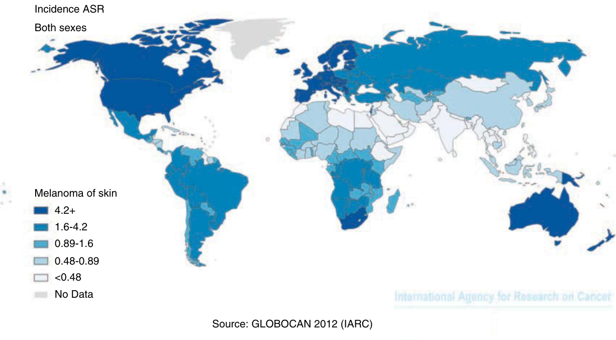
Figure 116-1 Estimated cancer incidence for melanoma world map. ASR = age-standardized rate. (Reproduced with permission from Ferlay J, Soerjomataram I, Ervik M, et al. GLOBOCAN 2012 v1.0, Cancer Incidence and Mortality Worldwide: IARC CancerBase No. 11 [Internet]. Lyon, France: International Agency for Research on Cancer; 2013. Available from: http:// globocan.iarc.fr, accessed 13 September 2016.)
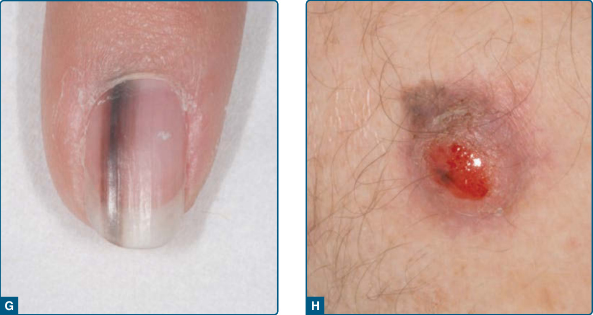
Figure 116-2 Clinical subtypes of melanoma: A, B, Superficial spreading melanoma, in A with extensive signs of regression (whole center of the lesion); C, ulcerated amelanotic nodular melanoma; D, pigmented nodular melanoma; E, lentigo maligna melanoma; F, acral lentiginous melanoma; G, subungual melanoma with melanonychia striata; H, ulcerated desmoplastic melanoma.
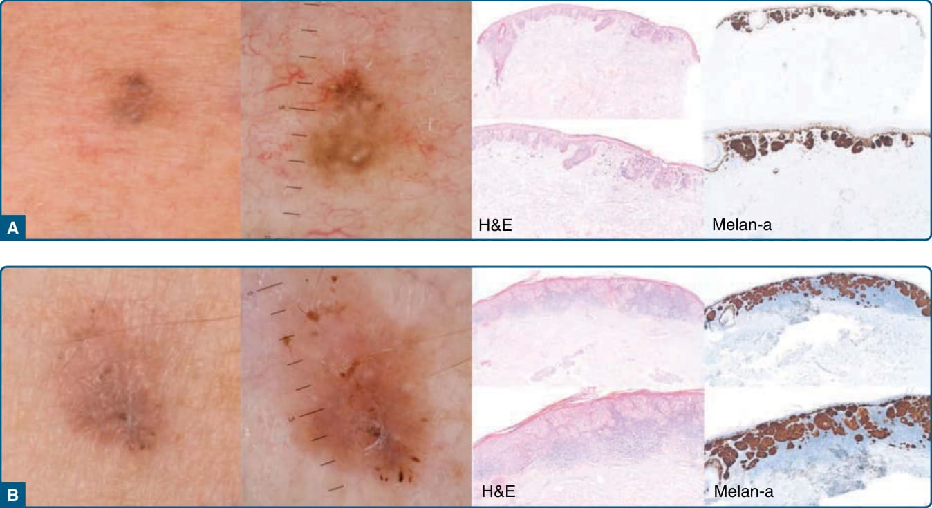
Figure 116-3 Clinical, dermoscopic and histologic presentation of 2 superficial spreading melanomas (A, B).
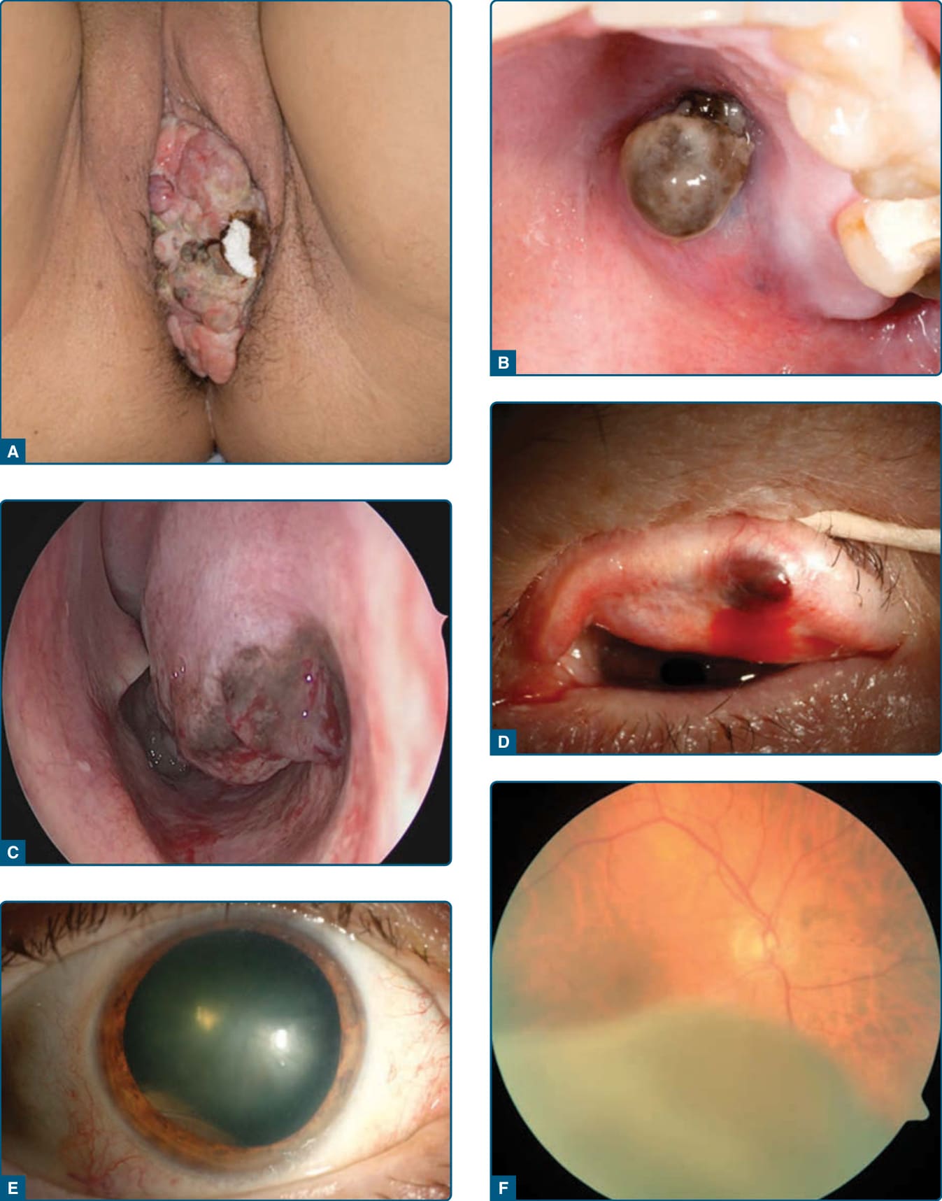
Figure 116-4 Mucosal melanomas: A, Introitus vaginae; B, gingiva; C, intranasal; D, conjunctiva; E, F uveal melanoma. (A-C, With friendly permission to publish from J. Thierauf, Department of ENT, University Hospital Heidelberg; D-F, Permission to publish from A. Scheuerle, Department of Ophthalmology, University Hospital Heidelberg.)
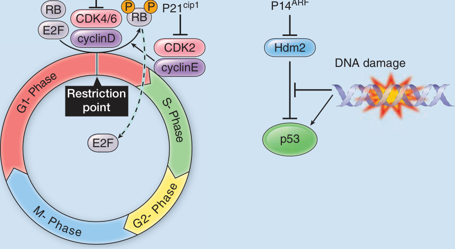
Figure 116-5 Mutations in the tumor suppressor gene CDKN2A account for approximately 40% of hereditary melanoma cases and lead to a loss of cell cycle control (A) and antiapoptosis (B).
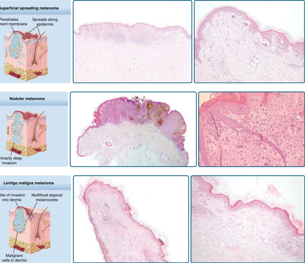
Figure 116-6 Histologic appearance (schematic, histopathologic overview, and close-up) of superficial spreading melanoma (A), nodular melanoma (B), and lentigo maligna melanoma (C).

Figure 116-7 Diagnostic algorithm.
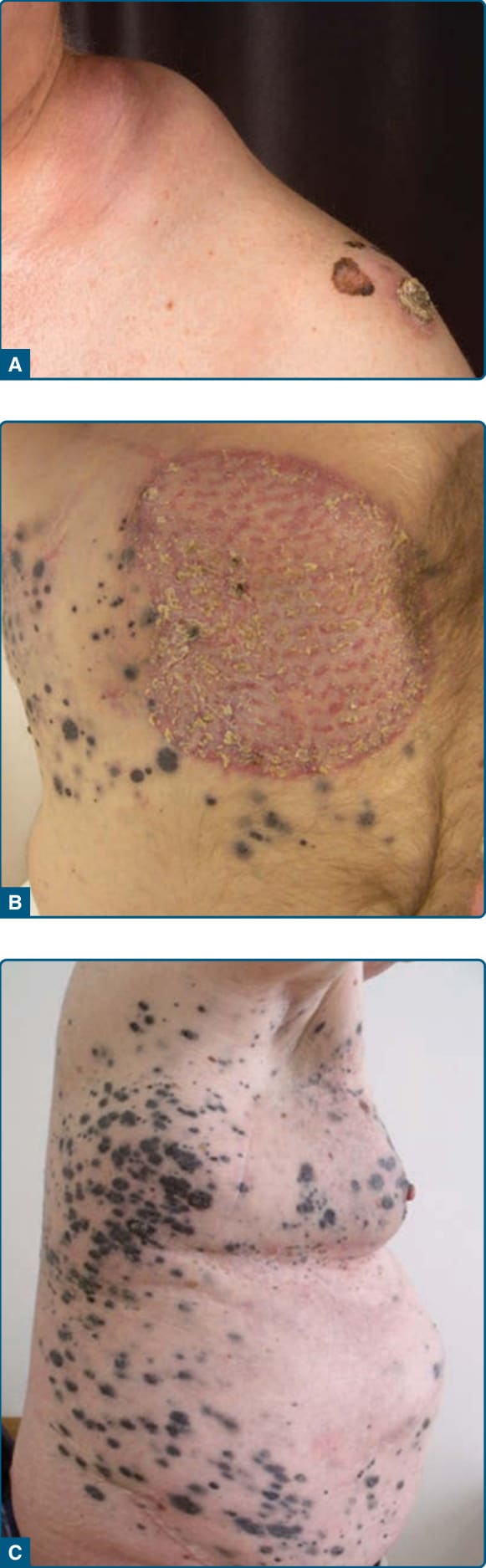
Figure 116-8 A, Patient with stage III disease (primary melanoma with bulky lymph node metastases). B, Patient with in-transit metastases. C, Patient with stage IV disease and extensive cutaneous metastases.
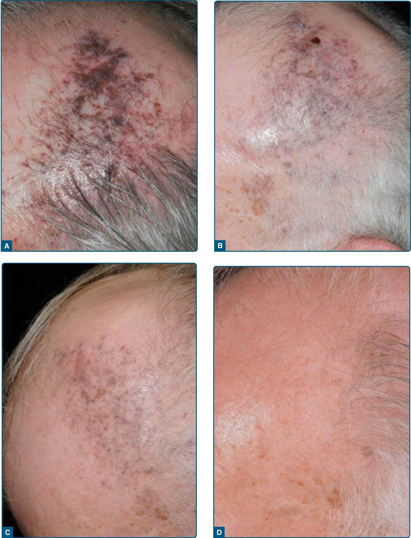
Figure 116-9 Treatment of satellite metastases with intralesional interleukin-2: A, before treatment; B, after 6 weeks of treatment; C, 4 weeks later without any further treatment; D, 2 years later without any further treatment.
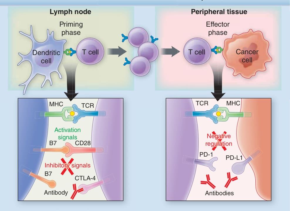
Figure 116-10 Mechanism of action of immune checkpoint blockage: The anti-CTLA-4 antibodies block the downregulation of T cells in their priming phase in the lymph nodes whereas the anti-PD-1 antibodies block the downregulation of activated T cells in their effector phase in the tumor microenvironment.
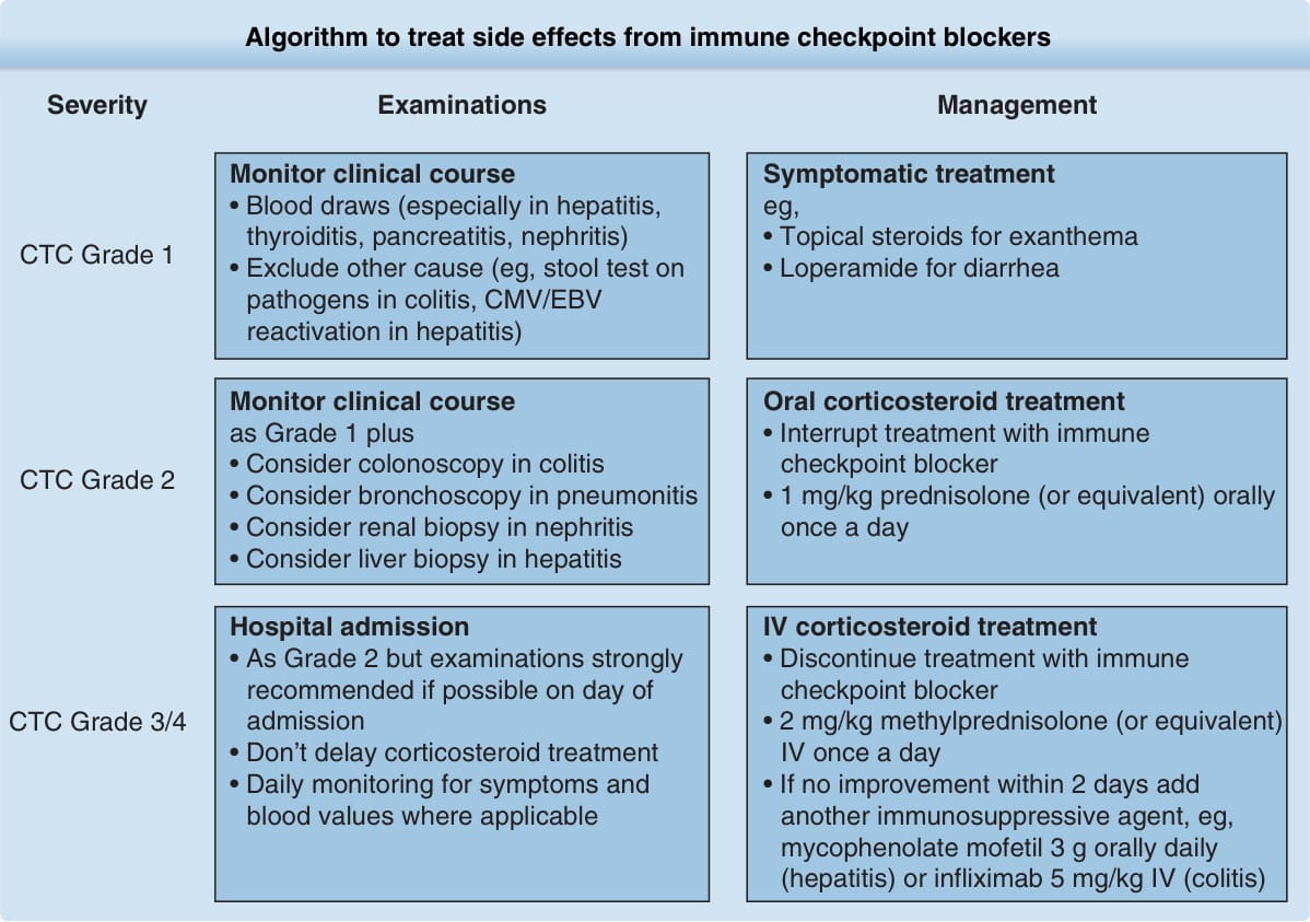
Figure 116-11 Algorithm to treat side effects from immune checkpoint blockers.
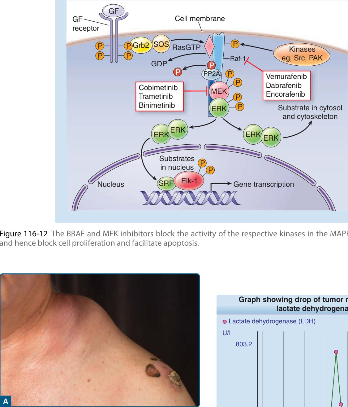
Figure 116-12 The BRAF and MEK inhibitors block the activity of the respective kinases in the MAPK signaling pathway and hence block cell proliferation and facilitate apoptosis.
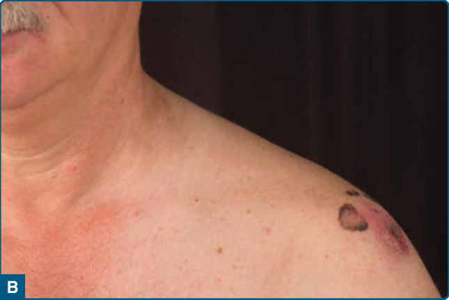
Figure 116-13 A 62-year-old patient with a superficial spreading melanoma on the left shoulder that metastasized to the regional lymph nodes already and was unresectable at the time of diagnosis (A). As a BRAF V600E mutation was detected, he received a combination treatment with the BRAF inhibitor dabrafenib and the MEK inhibitor trametinib. After 2 months of treatment, the lymph node conglomerates decreased in size significantly (B), accompanied by a drop of the tumor marker serum lactate dehydrogenase (C).
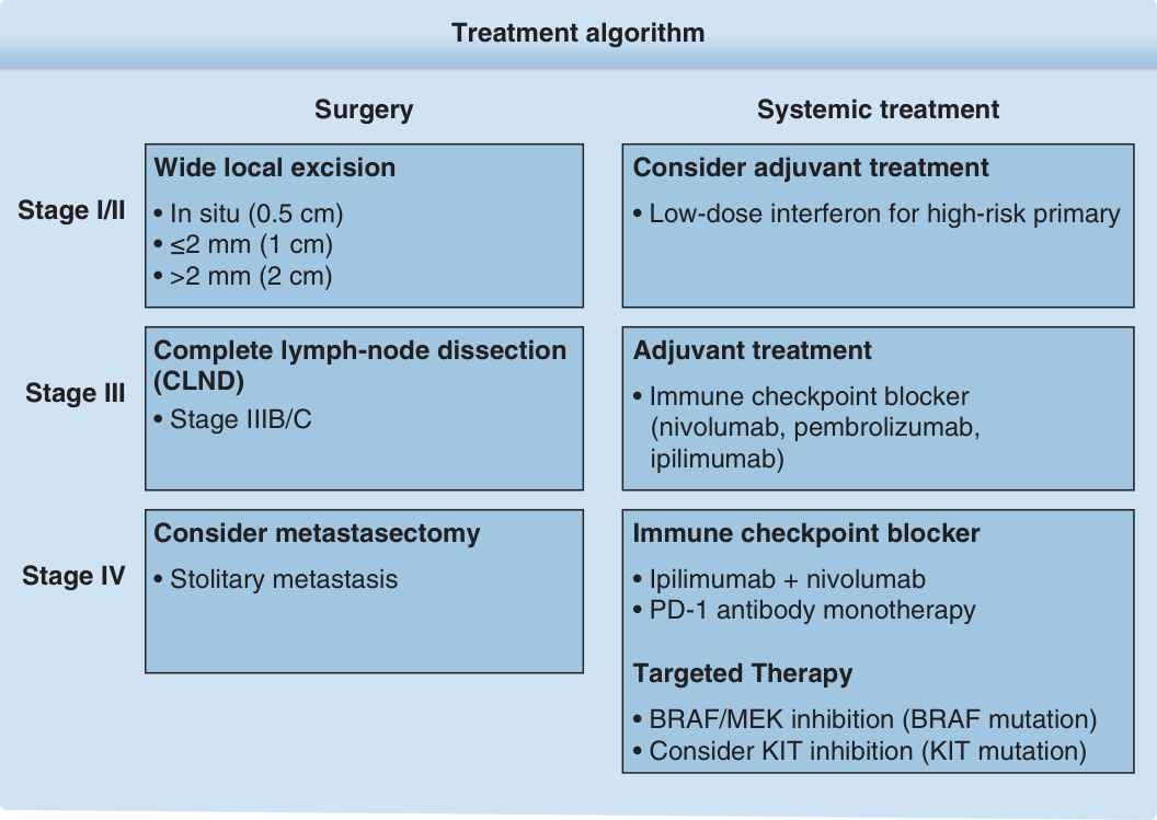
Figure 116-14 Treatment algorithm.
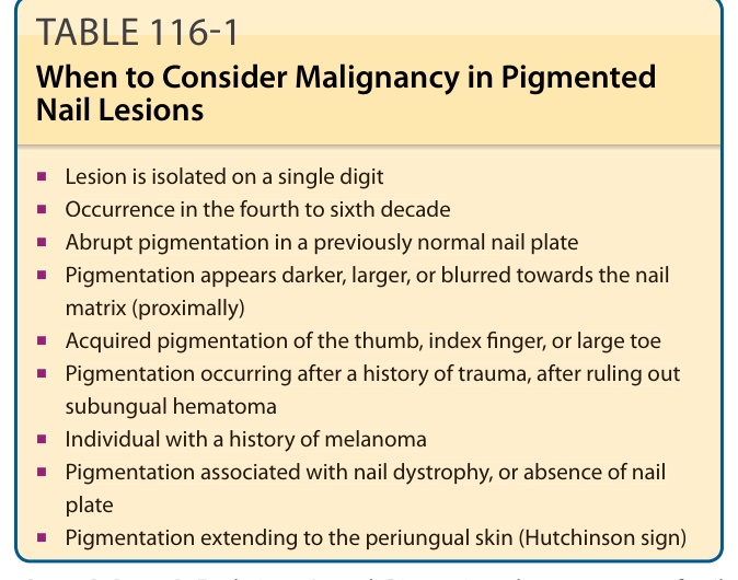
TABLE 116-1 When to Consider Malignancy in Pigmented Nail Lesions
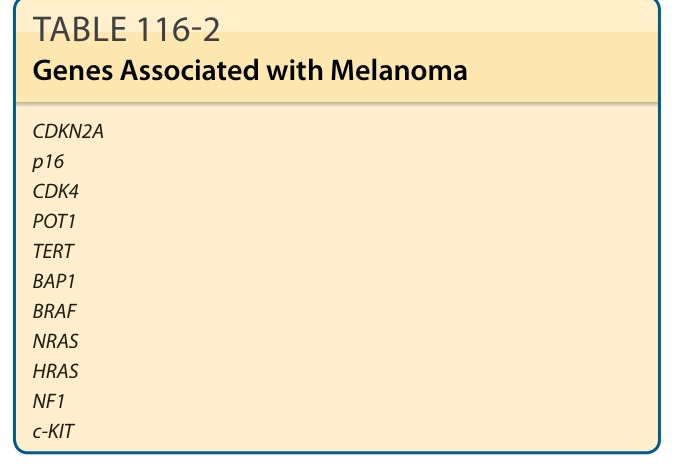
TABLE 116-2 Genes Associated with Melanoma

TABLE 116-3 ABCDE Checklista
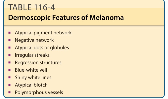
TABLE 116-4 Dermoscopic Features of Melanoma
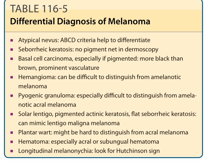
TABLE 116-5 Differential Diagnosis of Melanoma
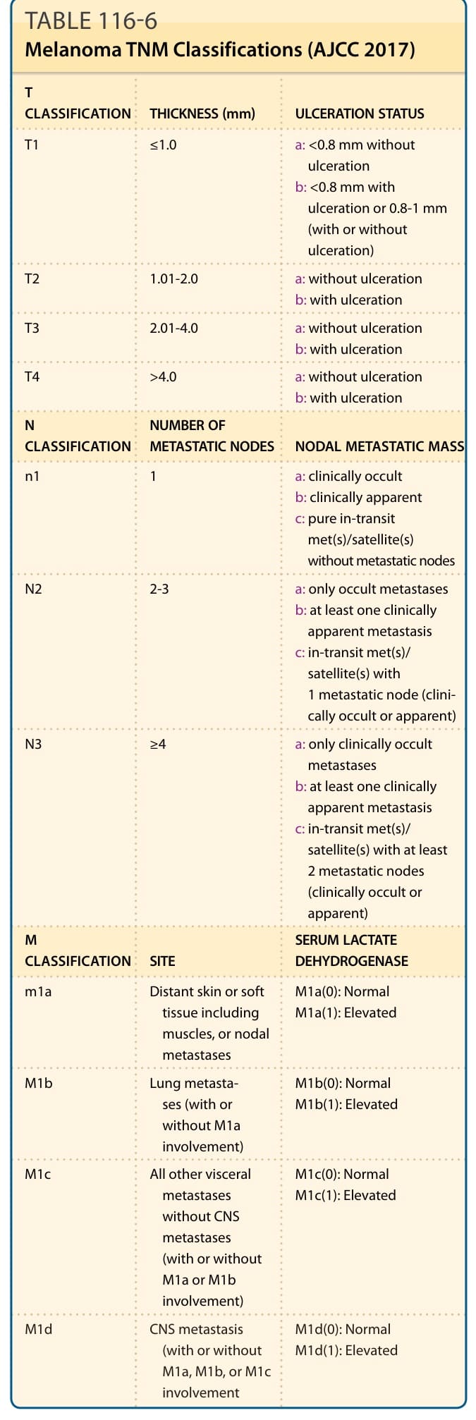
TABLE 116-6 Melanoma TNM Classifications (AJCC 2017)
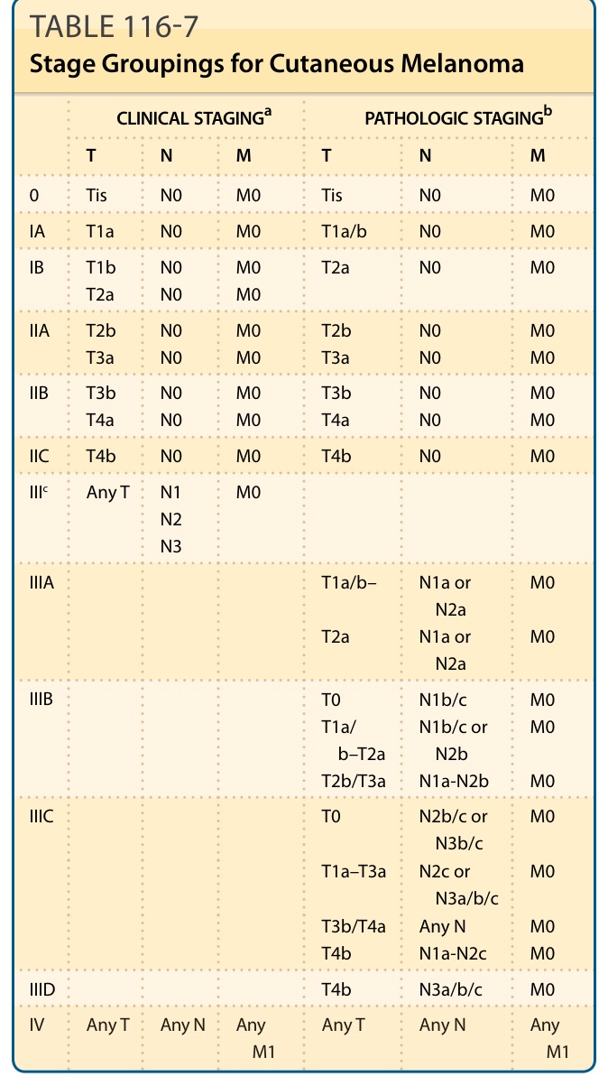
TABLE 116-7 Stage Groupings for Cutaneous Melanoma
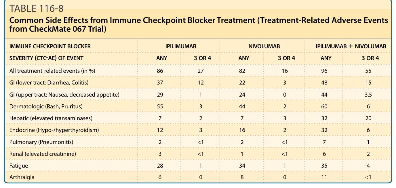
TABLE 116-8 Common Side Effects from Immune Checkpoint Blocker Treatment (Treatment-Related Adverse Events from CheckMate 067 Trial)
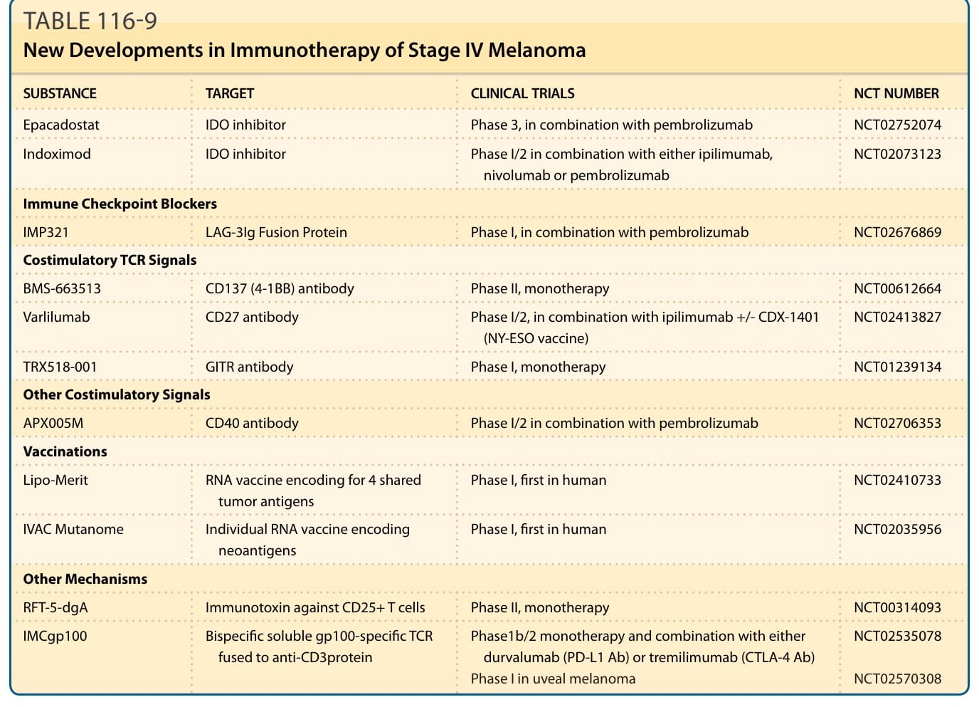
TABLE 116-9 New Developments in Immunotherapy of Stage IV Melanoma